Download
1 / 87
870 likes | 1.01k Views
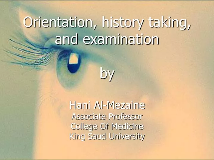
Orientation, history taking, and examination by Hani Al- Mezaine Associate Professor College Of Medicine King Saud University. Orientation. Course OPH 432. Objectives. a. To know the basic ophthalmic anatomy and physiology.

E N D
Orientation, history taking, and examinationbyHani Al-MezaineAssociate ProfessorCollege Of MedicineKing Saud University
Orientation Course OPH 432
Objectives a. To know the basic ophthalmic anatomy and physiology. b. To recognize assessment and management of common ophthalmic diseases. c. To know how to handle common ophthalmic emergencies. d. To handle simple ophthalmic diagnostic instruments. e. To be aware of common ophthalmic operations.
Components of the course • Lectures • Clinics • OR • Clinical sessions • ER
Lectures • History taking and physical examination in ophthalmology • Basic Anatomy and Physiology of the Eye • Lid, Lacrimal, and Orbit Disorders • Ocular emergencies and red eye • Strabismus, Amblyopia and Leukocoria • Acute Visual Loss
Chronic Visual Loss • Refractive Errors • Ocular manifestations of systemic diseases • Neuro-ophthalmology • Ocular Pharmacology and Toxicology
Clinical skill sessions • External Ocular Examination, Ocular motality and Alignment • Visual acuity and Ophthalmoscopy • Visual field, Tonometry, Pupill Examination
Marks distribution • 35 marks for the MCQs • 60 marks for the OSCE • 5 marks for attendance in clinical sessions, clinics, ER, OR.
Recommended textbooks 1. Required Text(s) a. Lecture notes Ophthalmology (latest edition) By: Bruce James (published by Blackwell Science) b. Basic Ophthalmology (latest edition) By: Cynthia A. Bradford (latest edition) (published by American Academy of Ophthalmology) c. Practical Ophthalmology: A manual for Beginning Residents (latest edition) By: Fred M. Wilson (published by American Academy of Ophthalmology
2. References • Vaughan and Asbury’s general Ophthalmology By: Paul Riordan-Eva (published by LANGE) • Clinical Ophthalmology: A Systematic Approach By : Jack T. Kanski (published by Butterworth Heinemann)
c. Electronic Materials, Web Sites 1. Department internet website. 2. Department’s teaching staff personal websites on University site. 3. University and KKUH/KAUH Library. 4. Audiovisual Unit of the Ophthalmology Department. 5. PubMed 5. Medscape 6. The digital journal of ophthalmology (www.djo.harvard.edu)
If there is a barrier between the examiner and the patient – a barrier with an opening of 1 square inch through which the examination can be performed. Which square inch you will choose??!!
Diagnostically and functionally, it is the most important square inch of the body surface.
Of all the organs of the body, the eye is most accessible to direct examination. • The external anatomy of the eye is visible to inspection with unaided eye and with fairly simple instruments. • The health or disease of a large portion of the body can be determined simply by looking at the eye.
The eye is so intimately connected with the rest of the body that it reveals enormous amount of general information. • Eye is the only part of the body where blood vessels and central nervous system tissues can be viewed directly.
The 12 cranial nerves provide us with a large part of our information about the brain. Of these , the eye examination evaluates CN II, III, IV, V, VI, VII, VIII. • In addition, it provides information about the autonomic pathways.(sympathetic /parasympathetic)
The best known connection between the brain and the eye is the ON. • The visual pathways, which extends from front to back across the brain can be studied easily and safely using perimetry. It can differentiates accurately between lesions of the temporal, parietal, and occipital lobes.
In addition, the ON has important clinical relationships to the pituitary gland, the middle ventricles, the venous sinuses, the meningeal and bony structures of base of the skull.
ON has the diagnostically useful capability of swelling with ↑ ICP (papilledema). Or visibly pale (optic atrophy) when its nerve fibers damaged at any point from Retina → LGB.
The study of CN III, IV, V, VI can evaluates the brain stem , cavernous sinus, apex of orbit .
Unilateral dilated pupil after head injury can occur due to pressure on pupil constrictor fibers of CN III. • CN VI involved in mastoid infection (petrous ridge) • Parotid gland, Inner ear disease → CNVII. • Nystagmus→ CN VIII
Focal brain lesions like: Vascular occlusions Hemorrhage Neoplasm Diffuse brain lesions like: Infections Demyelinating disorders → nerve damage.
Venous flow disorder: cavernous sinus thrombosis, carotid – cavernous fistula (orbital congestion)
Arterial emboli can reach the retina from carotid artery , heart valves, subacuteendocarditis. • Specific disease of the vessels like: PAN, temporal arteritis, HTN.
Hematological disorders of all types can manifest in the fundus. • Almost all metabolic disorders can affects the eye: DM:DR, cataract, RE, ophthalmoplegia. Hypothyroidism: cataract Wilson’s disease.
Thyroid eye disease: Exophthalmos, Lid retraction. Infections: (Syphilis,Toxoplasmosis, Rubella)
Mucocutaneous disorders: SJS, pemphigus Elastic tissue: (Pseudoxanthomaelasticum) Allergy: VKC
Chromosomal abnormalities: Trisomy: 13,15, 21. • The eye is a delicate indicator of poisoning: -Morphine addict → meiotic pupil -Lead poisoning, vitamin A intoxication → papilledema
I could go on and on about the discoveries possible in our most important square inch.‘’EYE’’ • 90% of our information reaches our brain via sight. • Unfortunately, of all the parts of the body, the eye is the most vulnerable to minor injury.
What are the objectives of the comprehensive ophthalmic evaluation?
Obtain an ocular and systemic history. • Determine the optical and health status of the eye and visual system. • Identify risk factors for ocular and systemic disease. • Detect and diagnose ocular diseases.
Establish and document the presence or absence of ocular symptoms and signs of systemic disease. • Discuss the nature of the findings and the implications with the patient. • Initiate an appropriate response. e.g. further diagnostic tests, treatment, or referral.
It is a gathering information process from the patient guided by an educated and active mind. • It is a selective guided and progressive elicitation and recognition of significant information • History by skilled person can arrive at the proper diagnosis in 90% of patients.
It gives vital guidance for: (a) physical examination (b) laboratory work (c) Therapy • Failure to take history can lead to missing vision or life threatening conditions.
Chief complaint: ’’The patient’s own words’’ ‘’she cannot see with the RE’’ You should not come to conclusion that her problem is nearsightedness and write down “Myopia of RE”. • The patient needs will not be satisfied until he/she has received an acceptable explanation of the meaning of the chief complaint and its proper management.
History of the Present Illness: • Detailed description of the chief complaint to understand the symptoms and course of the disorder. • Listen andquestion and then write down in orderly sequence that make sense to you.
* The time sequence when, How fast, what order did events occur? * Frequency, intermittency * location, Laterality * Severity * Associated symptoms * Documentation (old records, photo) e.gptosis, proptosis, VII N palsy. • gradual painless decrease vision both eyes for 1y. • Sudden painless decrease vision RE for 10 min.
“cannot see with RE”!! • ? Only distance vision blurred. • ? Blind spot is present in the center of VF • ? Right side of VF of the RE lost • ? Right VF of both eyes lost • ? A diffuse haze obscures the entire field of RE • Each of these has different diagnostic implication • Most pt. has difficulty providing precise and concise description
Disturbances of vision: • Blurred or decreased central vision • Decreased peripheral vision. (glaucoma) • Altered image size. (micropsia, macropsia, metamorphopsia). • Diplopia (monocular, binocular) • Floaters • Photopsia (flash of light)
Color vision abnormalities. • Dark adaptation problems. • Blindness (ocular, cortical). • Oscillopsia (shaking of images).
Ocular pain or discomfort: • Foreign body sensation • Ciliary pain (aching, severe pain in or around the eye, often radiating to the ipsilateral forehead, molar area) • Photophobia • Headache • Burning • Dryness • Itching: patient rub the eye vigorously (allergy) • Asthenopia(eye strain)
Abnormal ocular secretions: • Lacrimation, epiphora • Dryness • Discharge (purulent, mucopurulent, mucoid, watery)
Redness, opacities, masses • Anisocoria