Download
1 / 49
550 likes | 754 Views
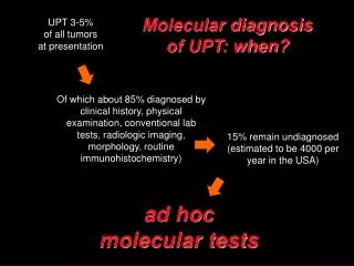
MOLECULAR DIAGNOSIS OF GENETIC EPILEPSY. Nagi ALHaj Prof of Molecular Biology Assist, Faculty Medicine , Sana’a ,University, Yemen Consultant of Genetic Center 48 MH. Overview on Genetic Epilepsy.

E N D
MOLECULAR DIAGNOSIS OF GENETIC EPILEPSY Nagi ALHaj Prof of Molecular Biology Assist, Faculty Medicine , Sana’a ,University, Yemen Consultant of Genetic Center 48 MH
Overview on Genetic Epilepsy A genetic contribution to etiology has been estimated to be present in about 40% of patients with epilepsy.. Three major groups: • Mendelian disorders, in which a single major locus can account for segregation of the disease trait • Non-mendelian or 'complex' diseases, in which the pattern of familial clustering can be accounted for by the interaction of the maternal inheritance pattern of mitochondrial DNA • Chromosomal disorders, in which a gross cytogenetic abnormality is present.
The ‘common’ non-familial idiopathic epilepsies tend to display ‘complex’ inheritance. • They including various forms of • Idiopathic generalised epilepsy (IGE) • Juvenile myoclonic epilepsy (JME) • Childhood absence epilepsy (CAE)
Genetics and Mutation • Mutations in over 2,000 genes have now been identified in patients with more than 3,000 different disease phenotypes. For the clinicians and their patients, it is becoming increasingly important to obtain a genetic diagnosis • Identifying the genetic aetiologyof a disease may influence clinical management and will provide information regarding risk to future pregnancies. • With the advent of high-throughput capillary sequencers and sequence analysis software, direct sequencing provides an accurate method for single gene analysis.
The inheritance pattern can be autosomal dominant, autosomal recessive, or X-linked. Mutations in a single gene may be associated with different types of seizures (clinical heterogeneity), and, conversely, mutations in different genes can cause the same epilepsy phenotype (genetic heterogeneity).
Traditional nomenclature of inherited epilepsy: Different mutations in different genes can result in different phenotypes Different mutations in different genes can result in similar phenotypes Different mutations within one gene can result in different phenotypes An identical mutation within one gene can result in different phenotypes in different individuals (cause: environment, other genes)
Mutation identification begins with a phenotype and proceeds toward the genotype genotype phenotype
The diagram shows the distribution of all genetic differences that had been mapped to chromosome 1 at the time this diagram was drawn.
mutation site Mutation identification by linkage analysis Mutational analysis can be used to identify cells or DNA that have genotype and allele frequency differences from the normal genome. • Genome scan has been replaced by mutational analysis but in a small number of families in whom the mutation cannot be identified • Remains the only method for the genetic diagnosis of carriers.
Mutant alleles generating defects in particular proteins could disrupt the dance . Cells homozygous for a mutant allele might be unable to complete chromosome duplication or mitosis or cytokinesis because a required component of the molecular machinery is missing or unable to function. A genetic map of part of the human X chromosome.
Elucidation of a disease mechanism presents a much more complex set of challenges cell protein network disease mechanism mRNA genotype phenotype
The number of potential defects increases exponentially with each emergent stage of complexity Not all potential defects arise from each mutation cell protein network disease mechanism mRNA genotype phenotype
The most effective target for therapy ( ) would be T the DNA mutation, but this is currently unfeasible Not all potential defects arise from each mutation cell protein network disease mechanism mRNA T genotype phenotype
The most effective target for therapy ( ) would be Proteins are also excellent targets for intervention T the DNA mutation, but this is currently unfeasible T cell protein network disease mechanism mRNA T genotype phenotype
The genetic contribution to epilepsy: the known and missing heritability. The Causes of Epilepsy, eds S.D. Shorvon et al, pp 6367. Cambridge University Press, Cambridge, 2011).
Genes and mutations in neonatal syndromes and GEFS+ KCNQ2 Chr. 20q13.3 Benign familial neonatal convulsions (BFNC); Benign neonatal epilepsy-1 (EBN1) BFNC/myokymia syndrome KCNQ3 Chr. 8q24 Benign familial neonatal convulsions (BFNC); Benign neonatal epilepsy-2 (EBN2)
SCN1B Chr. 19q13 Generalized epilepsy with febrile seizures plus, type 1 (GEFS+ type 1; GEFSP1) SCN1A Chr. 2q24 GEFS+ type 2; GEFSP2 Severe myoclonic epilepsy in infancy (SMEI) GABRG2 Chr. 5q33-q34 GEFS+ type 3; GEFSP3 SMEI SCN2A Chr. 2q23-q24 Febrile seizures associated with afebrile seizures Benign familial neonatal-infantile seizures (BFNIS)
Unknown Chr. 19q12-q13.1 Benign familial infantile convulsions, type 1 (BFIS type 1; BFIC1) Unknown Chr. 16p12-q12 BFIS type 2; BFIC2 Infantile convulsions and paroxysmal choreoathetosis (ICCA) Paroxysmal kinesigenic choreoathetosis (PKC) Unknown Chr. 16p12-p11.2 Rolandic epilepsy, paroxysmal exercise-induced dystonia, writer's cramp (RE-PED-WC) Unknown Chr. 16p13 Autosomal recessive (familial) benign idiopathic myoclonic epilepsy of infancy (FIME)
Mutations: E B A C A D B A F,G A C E E A C Gene 1 Gene 2 Gene 3 Gene 4 Gene 2 Gene 1 Gene 3 Gene 1 Gene 1 Gene 4 Gene 3 Gene 2 Gene 2 Traditional nomenclature of inherited epilepsy: Gene 1 Gene 2 Gene 3 Gene 4 Phenotype: Syndrome “A”: Syndrome “B”: Syndrome “C”: Syndrome “D”: Syndrome “E”: Syndrome “F”: Syndrome “G”: etc…
Gene-centric nomenclature of inherited epilepsy: Gene 1 Gene 2 Gene 3 Gene 4 Mutations: E B A C A D B A F,G A C E E A C Phenotype: Gene 1: Phenotypic range A, B, C, D Gene 2: Phenotypic range A, B, E, F, G Gene 3: Phenotypic range A, S, E Gene 4: Phenotypic range A, E etc… Both “Traditional” and “Gene-centric” nomenclatures have specific advantages and disadvantages
(1) The Infantile Epilepsy includes sequencing and deletion/duplication analysis of 38 genes causing Mendelianforms of epilepsy with onset of seizures during the first year of life. 38 gene activity, including • voltage-gated sodium channels, • the voltage-gated calcium channels, • and gamma-aminobutyricacid (GABAA) receptors.
(2) The Childhood-Onset Epilepsy 40 Genes (3)The Adolescent-Onset Epilepsy 21 Genes Includes sequencing and deletion/duplication analysis of 40 and 21 genes respectively that causing Mendelian forms of epilepsy. • Genes that encode nicotinic acetylcholine receptors and calcium channels,
Methodology • Infantile Epilepsy Panel • The Childhood-Onset Epilepsy • The Adolescent-Onset Epilepsy Using genomic DNA obtained from blood, ~ 570 coding exons and the flanking splice junctions of 38 genes are sequenced simultaneously by (next-generation sequencing). • The sequence is assembled and compared to published genomic reference sequences. • Sanger sequencing is used to compensate for low coverage and refractory amplifications in regions where pathogenic mutations have been previously published.
chromosome cell nucleus Double stranded DNA molecule Target Region for PCR Individual nucleotides DNA in the Cell
Extraction of DNA from whole Blood Extract and discard plasma, taking care not to remove the buffy coat.
5’ 3’ 5’ 3’ Starting DNA Template 3’ 5’ 3’ 5’ Separate strands (denature) Forward primer 5’ 3’ 3’ 5’ 5’ 3’ 5’ 3’ Make copies (extend primers) Reverse primer Add primers (anneal) DNA Amplification with the Polymerase Chain Reaction (PCR)
Original DNA target region Thermal cycle Thermal cycle Thermal cycle PCR Copies DNA Exponentially through Multiple Thermal Cycles In 32 cycles at 100% efficiency, 1.07 billion copies of targeted DNA region are created
M 1 2 3 4 5 6 7 8 9 10 11 loss of bands loss of bands Deletion analysis of the SCN1A gene by multiplex PCR.
Identification of a deletion encompassing gene in a male patient. Y-axes represent R Log ratio B allele frequency The red line (log R ratio profile) corresponds to the median smoothing series. The X-axis indicates the position on the X chromosome Identification of a hemizygous Xq22.1 deletion with a 370 K SNP microarray
Figure 1. Identification of a deletion encompassing PCDH19 in a male patient. Black horizontal bars represent the gene PCDH19) and pseudogenes comprised in the deleted region Analysis of the patient and his mother with CGH microarrays, showing that the new deletion occurred.
3’-TAAATGATTCC-5’ DNA template 5’ 3’ A Primer anneals AT Extension produces a series of ddNTP terminated products each one base different in length ATT ATTT ATTTA ATTTAC Each ddNTP is labeled with a different color fluorescent dye ATTTACT ATTTACTA ATTTACTAA ATTTACTAAG ATTTACTAAGG
Coriell Cell Repositories Human Gene Mutation Database Link to Gene OMIM Record Epilepsy GEFS
Links to Everywhere (almost) Gene Epilepsy GEFS
GEFS Genome Maps Gene NM records Gene Model UniGene NT Gene GEFS
Detection of 9 different point mutations of PCDH19 in 11 female patients by direct sequencing. The A of the ATG translation initiation codon in the reference sequence Sequence electropherograms of the mutations and the missense variant (c.3319C>G/p.Arg1107Gly) identified in association with the c.859G>T/p.Glu287X nonsense mutation.
Alignment of the regions surrounding the mutations (indicated by an arrow) in orthologous and paralogous proteins, showing the high conservation of each affected amino-acid in vertebrates and in the delta protocadherin paralogous genes.
FISH analysis of the gene deletion in the male patient showing somatic mosaicism in fibroblasts. A) Absence of the specific Xq22.1 probe site on metaphase chromosomes (PBL); (B) In fibroblasts, presence of one hybridization spot in 53% of the cells and absence of signal in the remaining 47%; FISH analysis on PBL (C) and fibroblasts (D) of a female control. PCDH19-specific signals (red) are indicated by arrowheads.