Download
1 / 35
460 likes | 1.08k Views
Preimplantation Genetic Diagnosis (PGD). Irene Souter, MD Director, PGD Program MGH Fertility Center. Pre-natal vs Pre-implantation diagnosis. Pre-natal Diagnosis. Amniocentesis Chorionic Villus Sampling (CVS). Pre-implantation Diagnosis. Introduced initially in 1990

E N D
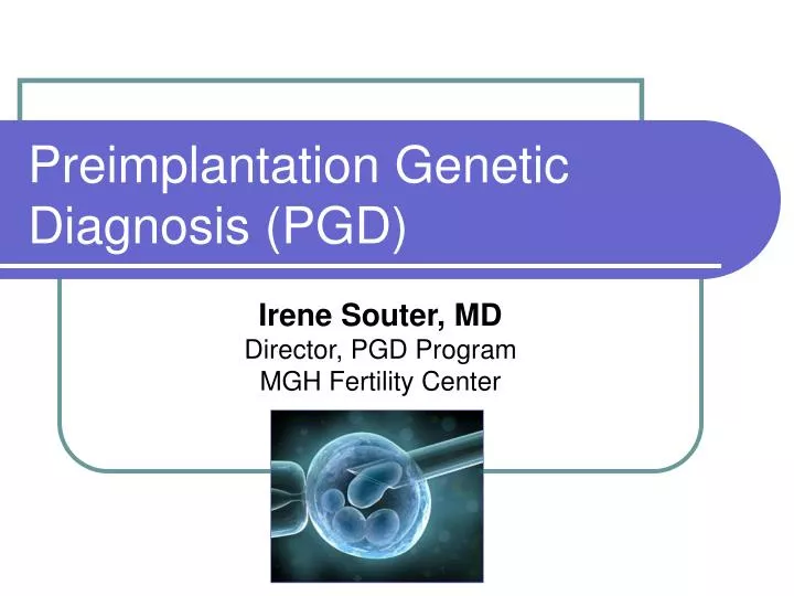
Preimplantation Genetic Diagnosis (PGD) Irene Souter, MD Director, PGD Program MGH Fertility Center
Pre-natal Diagnosis • Amniocentesis • Chorionic Villus Sampling (CVS)
Pre-implantation Diagnosis • Introduced initially in 1990 • Biopsy of a single cell per embryo, followed by its genetic diagnosis through different techniques (FISH, PCR, aCGH), and the subsequent replacement to the patient of those embryos classified by genetic diagnosis as normal.
PGD Indications Procedure is offered to couples: • With known single gene disorders that can be detected by PGD • With known chromosomal abnormalities that can be detected by PGD • requesting sex selection for X-linked disorders
PGD Indications • The procedure has also been offered to couples: undergoing IVF at risk for aneuploidy • maternal age > 35 yo • Prior trisomic conception • with recurrent pregnancy losses • Prior failed IVF cycles(>3 prior embryo transfers with high quality, morphologically normal embryos) • Requesting PGD for HLA-typing(to allow selection of embryos that are histocompatible with live siblings) • Requesting sex selection for “family balancing”
PGD Process • Ovulation Induction • Retrieval • Fertilization • Embryo Bx on Day-3 • Genetic Analysis • Embryo Transfer
Fertilization • Conventional Insemination • Intracytoplasmic Sperm Injection (ICSI)
Cleavage Stage Biopsy • Most widely used technique • Day 3, 6-8 cell stage
Genetic Analysis/PCR • DNA amplification • sequence harboring the mutation) • billions of copies in several hrs • Mutation Characterization • (by using mutation specific primers • by digestion with restriction enzymes • by heteroduplex analysis
ESHRE-Misdiagnosis • Single Gene Disorders • 14 cases (0.3%), 86% PCR • 8 babies born, 78% Prenatal Diagnosis • Translocations (FISH) • 3 cases, (0.08%) • No live births • PGS (FISH) • 10 cases, (0.08%) • One baby born with T-21 • SS • One case (0.2%), 46 XX, pregnancy terminated
Causes of Misdiagnosis • Human Error • Unprotected sex • mislabeling, misidentification, misinterpretation • wrong embryo transfer • incorrect probes or primers • Technical • Probe or primer failure • contamination (maternal, paternal, operator, carry-over) • Intrinsic (embryo) • Mosaicism • Allele drop out • Uniparental Disomy
ESHRE-Delivery Outcomes • 3163 deliveries • 3841 children born • 24% multiples, 96% twins • 28% preterm births • 17% singletons • 67% twins • 74% triplets • Mean Birth Weight: • 3215 grs • 2400 grs (twins) • Mean Length: 50.0 cm
PGD & Malformations ESHRE PGD Consortium, 2003 Major malformations: 2.6% • Phocomelia and pulmonary deficiency, chylothorax, congenital hip dislocation, abdominal cystic mass, pes equinivarus, exencephaly Minor malformations: 1.4% • syndactyly, hydrocele testis, ASD, mongolian spot, sacral dimple Liebaers et al, Belgium 2010 Major malformations: 2.1% vs ICSI: 3.4% • chylothorax, VSD, oeasophageal atresia, cataract, umbilical hernia, ichthyosis, cardiopathy
ESHRE-Single Gene Disorders • Most common autosomal recessive disorder • -thalassemia/sickle cell anemia, CF, SMA • Most common autosomal dominant disorder • Myotonic Dystrophy, Huntington Disease, NF-1, Charcot-Marie-Tooth • Most common X-linked disorder • Fragile X, DMD, and Becker Muscular Dystrophy • Hemophilia A and B • 3530 Cycles, dx: 85.8% of the biopsied embryos • CPR: 23% per OR, 29.5% per ET
Conclusions • For couples at risk for producing offspring with either debilitating monogenic disorders or chromosomal abnormalities IVF/PGD represents a major scientific advance.
Conclusions • Complications, both before and after birth, are no different in type or number from those found in a comparable ICSI population • Other parameters such as birth weight and length, are also similar to an ICSI population • PGD appears to be a safe method to avoid the birth of children with genetic defects
Conclusions • Before PGD is performed, genetic counseling must be provided to ensure that patients fully understand the • risk for having an affected child • the impact of the disease • the available options • the multiple technical limitations including the possibility of an erroneous result • Prenatal diagnostic testing is strongly encouraged to confirm the results of PGD
What’s in the Future? • With the advent of the microarray techniques for the analysis of the genome, transcripts of thousands of genes can be tested at one time, and the combination of both might dramatically change our future