Download
1 / 104
1.11k likes | 1.71k Views
Topic 3 Autoimmunity. Terry Kotrla, MS, MT(ASCP)BB Fall 2005. Introduction. Under normal circumstances immune system will not destroy self antigens.

E N D
Topic 3 Autoimmunity Terry Kotrla, MS, MT(ASCP)BB Fall 2005
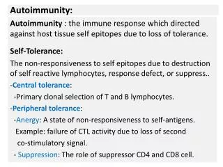
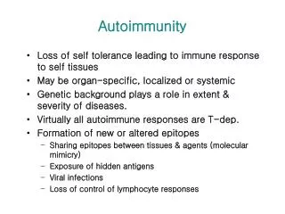
Introduction • Under normal circumstances immune system will not destroy self antigens. • Autoimmunity can be defined as breakdown of mechanisms responsible for self tolerance and induction of an immune response against components of the self. • In numerous autoimmune diseases it is well recognized that products of the immune system cause damage to the self.
Autoimmune Response • Antibody directed against “self”, termed auto-antibody • Considered abnormal but usually does not result in disease. • May occur in healthy individuals.
Autoimmune Disease • Disorder in which tissue injury is caused by an immunologic reaction of the host to its own tissues. • Precise mechanisms unknown. • Classified as systemic or organ specific, frequently have overlap.
Proposed Mechanisms • Forbidden clone • Altered antigen • Sequestered Antigen • Immunologic deficiency theory • Genetic influence
Forbidden clone • Clone of changed or altered lymphocytes arise through mutation. • Lack foreign surface antigens, not destroyed. • Because of alteration may recognize host as foreign.
Altered Antigen • Surface antigens on host altered by chemical, biological or physical means. • This new antigenic determinant may be recognized as foreign by the host.
Sequestered Antigen • Some antigens in the body are hidden from cells of the immune system. • If there is damage to these organs causing exposure of these sequestered antigens an immune reaction to these antigens may occur.
Immunologic Deficiency Theory • Relates the increased frequency of auto-antibodies and increased immune system deficiency to age. • Mutation or loss of immune regulatory powers results in the condition in which self antigens behave as foreign antigens.
Genetic Influence • It is well recognized that certain immune disorders predominate in females and in families. • Determined by family studies. • Genetic links have occurred between diseases and HLA antigens
Contributing Factors • Defects in the immune system. • Influence of hormones • Environmental conditions
Classification of Autoimmune Diseases • Systemic- the auto-immunity is directed against an antigen that is present at many different sites and can include involvement of several organs • Organ specific - Organ specific means the auto-immunity is directed against a component of one particular type of organ. • Both – can get overlap
Systemic Lupus Erythematosus • Chronic, systemic inflammatory disease caused by immune complex formation. • The word "systemic" means the disease can affect many parts of the body. • Pathophysiology associated with clinical features secondary to immune complexes depositing in tissues resulting in inflammation. • Parts of the body affected include: the joints, skin, kidneys, heart, lungs, blood vessels, and brain.
Systemic Lupus Erythematosus • Peak age of onset is 20 to 40 years of age. • Found more frequently in women. • Has both genetic and environmental factors.
SLE Clinical Signs • Extremely diverse and nonspecific. • Joint involvement most frequent sign: polyarthralgia and arthritis occur in 90% of patients. • Skin manifestations next most common. • Erythematosus rash may appear. • Most classic is butterfly rash.
SLE Butterfly Rash • The source of the name "lupus" is unclear. All explanations originate with the characteristic butterfly-shaped malar rash that the disease classically exhibits across the nose and cheeks. • In various accounts, some doctors thought the rash resembled a wolf pattern. In other accounts doctors thought that the rash, which was often more severe in earlier centuries, created lesions that resembled wolf bites or scratches. • Stranger still, is the account that the term "Lupus" didn't come from latin at all, but from the term for a French style of mask which women reportedly wore to conceal the rash on their faces
SLE Clinical Signs • Renal involvement very common. • Caused by deposition of immune complexes in kidney tissue. • Leads to renal failure, most common cause of death. • Other systemic effects: • Cardiac • Central nervous system. • Hematologic abnormalities.
Immunologic Findings • Lupus Erythematosus (LE) cell, neutrophil which has engulfed the antibody-coated nucleus of another cell. • First classic test to aid in diagnosis. • Not utilized anymore, may still see in older references. • Over activity of B cells main immunologic characteristic. • Antinuclear antibodies produced. • More than 28 antibodies associated with LE have been identified. • Level of antibody production correlates with severity of symptoms. • Estrogen enhance B cell activation.
LE Cell • Here is the famous "LE cell" test which has value only in demonstrating how the concept of autoantibodies work. The pink blobs are denatured nuclei. Here are two, with one seen being phagocytozed in the center by a PMN. This test is not nearly as sensitive as the ANA which has supplanted the LE cell test. Therefore, NEVER order an LE cell test. [Image contributed by Elizabeth Hammond, MD, University of Utah]
Immunologic Findings • Decrease in absolute number of T cells • Accumulation of immune complexes with activation of complement lead to kidneydamage. • Drug induced lupus may occur, discontinue drug, symptoms usually disappear.
Laboratory Diagnosis • Screening test for anti-nuclear antibodies (ANA) first test done. • Antibodies directed against nuclear material of cells. • Flourescent anti-nuclear antibody (FANA) most widely used, extremely sensitive, low diagnostic specificity. • Animal or human cells fixed to slide. • Add patient serum and incubate. • Wash to remove unreacted antibody. • Add anti-human globulin labeled with fluorescent tag or enzyme.
ANA • Patterns of reactivity: • Homogenous-entire nucleus stained • Peripheral-rim of nucleus stained • Speckled-spots of stain throughout nucleus • Nucleolar-nucleolus only stained • False positives and negatives occur. • If positive, perform profile testing.
Antinuclear Antibody Test • Antinuclear antibodies (ANA) are autoantibodies against various cell nucleus antigens and are found in patients with autoimmune diseases such as SLE. • Some of ANA are considered to be useful for diagnosis of autoimmune diseases.
Homogeneous Pattern • Smooth, even staining of the nucleus with or without apparent masking of the nucleoli
Nucleolar • 23 or 46 (or some multiple of 46) bright speckles or ovoid granules spread over the nucleus of interphase cells
Peripheral • Fluorescence is most intense at the periphery of the nucleus with a large ring starting from the internal nuclear membrane and the rest of the nucleus showing weaker yet smooth staining.
Speckled • Large speckles covering the whole nucleoplasm, interconnected by a fine fluorescent network.
Anti-nuclear antibodies detected by FANA • Double-stranded DNA (ds-DNA) antibodies are most specific for SLE, correlate well with disease activity. • Antihistone antibody second major antibody found in SLE. • Deoxyribonucleoprotein (DNP) antibody, responsible for LE cell phenomena and available as a latex agglutination test. • Anti-Sm antibody, specific for LE. • SS-A/Ro and SS-B/La antibodies, most common in patients with cutaneous manifestations. • Anti-nRNP detected in patients with SLE as well as mixed connective tissue disease. • Presence of antibodies not diagnostic, may be present due to other diseases.
Anti-nuclear Antibodies by Immunodiffusion. • Used to determine specificity. • Ouchterlony double diffusion most frequently used to identify antibodies to: Sm, nRNP, SS-A/Ro, SS-B/La and others. • Test is not as sensitive but very specific.
Extractable Nuclear Antigen • This is antibody to a cytoplasmic ribonuclear protein complex. • It is associated with mixed connective disease and SLE with particular features (arthritis, myositis, Raynaud's phenomenon - also association with HLA-DR4 and HLA-DQw8).
Antiphospholipid Antibodies • Antiphospholipid antibodies may be present and are of two types. • Anticardiolipin. • Lupus anticoagulant, if present, may cause spontaneous abortion and increase • Risk of clotting, platelet function may be affected.
Treatment • Aspirin and anti-inflammatories for fever and arthritis. • Skin manifestations-anti-malarials or topical steroids. • Systemic corticosteroids for acute fulminant lupus, lupus nephritis or central nervous system complications. • Five year survival rate is 80 to 90%.
Rheumatoid Arthritis • Chronic inflammatory disease primarily affecting the joints, but can affect heart, lung and blood vessels. • Women three more times as likely as men to have it. • Typically strikes at ages between 20 and 40, but can occur at any age. • The three major symptoms of arthritis are joint pain, inflammation, and stiffness. • Progress of disease varies.
Clinical Signs • Diagnosis based on criteria established by American College of Rheumatologists, must have at least 4 of the following: • Morning stiffness lasting 1 hour. • Swelling of soft tissue around 3 or more joints. • Swelling of hand/wrist joints. • Symmetric arthritis.Subcutaneous nodules • Positive test for rheumatoid factor. • Xray evidence of joint erosion.
Clinical Signs • Symptoms initially non-specific: malaise, fever, weight loss, and transient joint pain. • Morning stiffness and joint pain improve during the day. • Symmetric joint pain: knees, hips, elbows, shoulders. • Joint pain leads to muscle spasm, limits range of motion, results in deformity. • Approximately 25% of patients have nodules over bones (necrotic areas), nodules can also be found in organs. • Certain bacteria may trigger RA due to certain proteins that possess antigens similar to those antigens found in joint, ie, molecular mimicry
Immunologic Findings • Rheumatoid Factor (RF) is an IgM antibody directed against the Fc portion of the IgG molecule, it is an anti-antibody. • Not specific for RA, found in other diseases. • Immune complexes form and activate complement and the inflammatory response. • Enzymatic destruction of cartilage is followed by abnormal growth of synovial cells, results in the formation of a pannus layer.
Diagnosis • Diagnosis is based on: • Clinical findings. • Radiographic findings • Laboratory testing. • Laboratory tests involve testing patients serum with red blood cells or latex particles coated with IgG, agglutination is a positive result. • Nephelometry and ELISA techniques are available to quantitate the RF. • Erythrocyte Sedimentation Rate (ESR) used to monitor inflammation. • C-Reactive protein (CRP) is utilized to monitor inflammation
Treatment • Rest and nonsteroidal anti-inflammatory drugs control swelling and pain. • Substantial functional loss seen in 50% of patients within 5 years. • Slow acting antirheumatic drugs are coming into use but have side affects. • Joint replacement.
Hashimoto's Thyroiditis • Hashimoto's Thyroiditis is a type of autoimmune thyroid disease in which the immune system attacks and destroys the thyroid gland. • The thyroid helps set the rate of metabolism - the rate at which the body uses energy. • Hashimoto’s prevents the gland from producing enough thyroid hormones for the body to work correctly. • It is the most common form of Hypothyroidism (underactive thyroid).
Hashimoto’s Thyroiditis • Organ specific disease affecting the thyroid gland. • Most often seen in women 30 to 40 years old, may be genetic predisposition. • Common cause of hypothyroidism. • Causes diffuse hyperplasia in the gland resulting in development of a goiter. • Thyroid autoantibodies are formed.
Hashimoto’s Thyroiditis • Hashimoto's thyroiditis is the most common cause of hypothyroidism. • It is also most prevalent in elderly women and tends to run in families. • Hashimoto's thyroiditis occurs eight times more often in women than men. • Certain chromosomal abnormalities include Hashimoto's thyroiditis as a symptom.
Symptoms • The following are the most common symptoms. However, each individual may experience symptoms differently: • goiter (enlarged thyroid gland which may cause a bulge in the neck) • other endocrine disorders such as diabetes, an underactive adrenal gland, underactive parathyroid glands, and other autoimmune disorders • fatigue • muscle weakness • weight gain
Thyroid • Thyroid hormones are produced by the thyroid gland. This gland is located in the lower part of the neck, below the Adam's apple. • The gland wraps around the windpipe (trachea) and has a shape that is similar to a butterfly - formed by two wings (lobes) and attached by a middle part (isthmus).
Goiter • This enlargement is due to the inflammatory cells which destroy thyroid cells, resulting in long term scarring. When the cells are damaged they cease thyroid hormone production, resulting in hypothyroidism • A goiter only needs to be treated if it is causing symptoms. • The enlarged thyroid can be treated with radioactive iodine to shrink the gland or with surgical removal of part or all of the gland (thyroidectomy). • Small doses of iodine (Lugol's or potassium iodine solution) may help when the goiter is due to iodine deficiency.
Laboratory Testing • The diagnosis of Hashimoto's thyroiditis is simply diagnosed by two blood tests. • Routine thyroid function tests to confirm that a patient has an underactive thyroid gland. • Anti-microsomal and anti-thyroglobulin antibodies are immune cells which the body produces to attack specific portions of the thyroid cells which pinpoint Hashimoto's thyroiditis as the cause of the hypothyroidism. • The anti-microsomal antibody test is much more sensitive than the anti-thyroglobulin, therefore some doctors use only the former blood test. • These thyroid autoantibodies blood tests are high in about 95% of patients with Hashimoto's thyroiditis, but are not diagnostic.