Download

1 / 83
840 likes | 1.02k Views
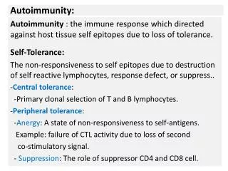
AUTOIMMUNITY. Johan van Rensburg. What goes wrong?. Tissue damage due to antibodies T cells Etiology multi-factorial environmental genetic specific HLA genotypes. Self antigens. HLA II > HLA I. Tolerance. Tolerance to self-antigens deletion of self-reactive B and T cells

E N D
AUTOIMMUNITY Johan van Rensburg
What goes wrong? • Tissue damage due to • antibodies • T cells • Etiology multi-factorial • environmental • genetic • specific HLA genotypes Self antigens HLA II > HLA I
Tolerance • Tolerance to self-antigens • deletion of self-reactive B and T cells • during maturation in the bone marrow and thymus • Recognition of peptide/MHC complexes by peripheral T cells • In the absence of costimulatory signals No activation of potentially self-reactive T cells Anergy
Tolerance • Regulatory(suppressor) T cells • Immunologically privileged sites • eye • brain TGFB IL-10 IL-4 Clonal expansion of autoreactive cells Do not normally encounter immunocompotent cells Self antigen inaccessible or in to low quantities
Autoimmune disease • Lack of tolerance • Triggered by • Infection • Other environmental factors • Unknown Increased/ aberrant expression of costimulatory molecules Changing antigenicity of infected tissue (now a target) Molecularmimicracy(eg. Rheumatic fever) Superantigens Stimulates families of T cells expressing a particular TCR-Vß segment
GENETIC PREDISPOSITION MHC-class II genes Other MHCgenes Multiple non-MHC genes Environmental factors CD4+ T cell driving force (autoreactive) Autoreactive B cells CD8+ T-cells IgG autoantibodies Non T cell effector cells Cell-mediated organ damage Autoantibody mediated organ damage
In general • Most common of multisystem connective tissue disease • Geographical • Racial • Caucasians • 30/100 000 • Afro-caribians • 200/100 000 • Gender • 90% Women • Age • Peak • 2nd to 3rd decades
Etiology and pathogenesis • Polyclonal B and T cell activation • Circulating auto-antibodies • Multi-organ involvement • Wide variety of clinical presentation • ~50 autoantigens identified • Hidden from Immune system in health • Intranuclear • Intra-cellular • Possible mechanisms • Environmental factors • Apoptosis • Autoantigens expressed on cell surface • Induce flares of SLE • Pregnancy • Infection • Sunlight
Diagnosis • Revised ACR criteria for SLE • 4/11 in present or past • 4 mucocutaneous • 4 Systems • 1 pleura/pericardial/peritoneum (PPP) • 2 Auto-antibody • Anti-dsDNA • 30-50% of patients • ANA negative unlikely for SLE • Unless (Ro) positive • Most have skin rashes
4 mucocutaneous • Malarrash • Erythema • Fixed • Flat • Raised • Sparing nasolabial folds • Discoid rash • Erythematous • Raised patches • Adherent • Keratotic scaring • Follicular plugging • Photosensitivity • Skin rash • Sun burn areas • Oral ulcers • May be painless
4 Systems • Arthritis/arthralgia • Non-erosive • 2 or more peripheral joints • (Jaccoudarthropathy) • Renal • Proteinuria • Persistent • >0.5g/day • Cellular casts • Red cell • Granular • Tubular • Neurological • Seizures • Psychosis • Absence • Offending drugs • Metabolic derangement • Hematological • In absence of offending drugs • Hemolytic anemia • Leucopenia • <4000/mm3 • 2 separate occasions • Lymphopenia • <1500/mm3 • 2 separate occasions • Thrombocytopenia • <100 000/mm3 • 2 Separate occasions
JACCOUD ARTHRITIS Jaccoud Sigismond, French physician, 1830–1913
GLOMERULONEPHRITIS • Minimal change • Mesangeal • Focal proliferative • Diffuse proliferative • Membranous glomerulonephritis • End Stage renal failure • (Typical is that the complement is diminished)
1 pleura/pericardial/peritoneum (PPP) • Serositis • Pleuritis • Pleuritic pain • Rub • Effusion • Pericarditis • ECG • Rub • Effusion
2 Auto-antibody • Immunology • Anti-DNA antibodies • Anti-SM antibodies • Antiphosfolipid antibodies • Antinuclear antibodies (ANA) • Abnormal titer • By immunofluorescence
Clinical features(more than criteria) • Raynaud's phenomenon • Musculoskeletal • Mucocutaneous • Renal • Management • Cardiopulmonary • Central nervous system • Hematological • Other
Classiffication • Adult polymiositis • Adult dermatomyositis • Important • Malignancy • Childhood dermatomyositis
Clinical picture • Proximal muscle weakness • Skin manifestations • NOT typically sun exposed areas • Malar rash • Heliotropic rash • V-sign • Shawel sign • Gottron’s nodules • Mechanic hands
MALAR AND HELIOTROPIC RASH Upper eyelid Involving nasolabial folds
Differential diagnosis (NB) • Inclusion body myositis • Other causes of proximal muscle weakness • See Davidson’s
Clinical features • Risk markers • Common clinical features • Less common features • Auto antibodies • Associated autoimmune disorders
Scleroderma (systemic sclerosis) Scleros: hard Derma: skin
SYSTEMIC SCLEROSIS Multisystem disorder Fibrosis of the skin, bloodvessels, and viseral organs Affected organs: Lungs Heart Kidneys GIT
CLASSIFICATION • Limitedcutaneous disease • Diffusecutaneous disease • Sinescleroderma • Undifferentiated connective tissue disease • Overlapsyndromes
Limited cutaneous scleroderma • Skin thickening: Distal to elbow and knee Face and neck • Synonym: CREST Syndrome
Diffuse cutaneous scleroderma • Skin thickening includes: Trunk Face Proximal extremities Distal extremities
Sine scleroderma • Internal organ manifestations • Vascular abnormalities • Serologic abnormalities • Noclinical detectable skin change