Download
1 / 10
100 likes | 104 Views
Here in this paper we discuss about an efficient method k means clustering for detection of tumour volume in brain MRI scans. This paper describes an efficient method for automatic brain tumor segmentation for the extraction of tumour tissues from MR images. It combines Perona and Malik anisotropic diffusion model for image enhancement and K means clustering techniques for grouping tissues belonging to a specific group. The developments in the application of information technology have completely changed the world. The obvious reason for the introduction of computer system is reliability, accuracy, simplicity and ease of use. Besides, the customization and optimization features of a computer system and among the other major driving forces in adopting and subsequently strengthening the computer aided systems. On medical imaging, an image is captured, digitized and processed fordoing segmentation and for extracting important information. Manual segmentation is an alternate method for segmenting an image. This method is not only tedious and time consuming, but also produces inaccurate results. Therefore, there is a strong need to have some efficient computer based system that accurately defines the boundaries of brain tissues along with minimizing the chances of user interaction with the system. Ananthagiri Vijaya Saradhi | L. Srinivas "An Efficient Method K-Means Clustering for Detection of Tumour Volume in Brain MRI Scans" Published in International Journal of Trend in Scientific Research and Development (ijtsrd), ISSN: 2456-6470, Volume-2 | Issue-4 , June 2018, URL: https://www.ijtsrd.com/papers/ijtsrd13027.pdf Paper URL: http://www.ijtsrd.com/engineering/electronics-and-communication-engineering/13027/an-efficient-method-k-means-clustering-for-detection-of-tumour-volume-in-brain-mri-scans/ananthagiri-vijaya-saradhi<br>

E N D
International Research Research and Development (IJTSRD) International Open Access Journal t Method K-Means Clustering for Detection of Tumour Volume in Brain MRI Scans Detection of Tumour Volume in Brain MRI Scans International Journal of Trend in Scientific Scientific (IJTSRD) International Open Access Journal ISSN No: 2456 ISSN No: 2456 - 6470 | www.ijtsrd.com | Volume 6470 | www.ijtsrd.com | Volume - 2 | Issue – 4 An Efficient Method K Detection of Tumour Volume in Brain MRI Scans Means Clustering for Ananthagiri Vijaya Saradhi M.Tech Scolar, Digital Image Processing, Nalanda Institute of Engineering and Technology (NIET), Guntur District, Andhra Pradesh, India ABSTRACT Here in this paper we discuss about an efficient method k-means clustering for detection of tumour volume in brain MRI scans. This paper describes an efficient method for segmentation for the extraction of tumour tissues from MR images. It combines Perona and Malik anisotropic diffusion model for image enhancement and K means clustering techniques for grouping tissues belonging to a specific group. The developments in the application of informa technology have completely changed the world. The obvious reason for the introduction of computer system is: reliability, accuracy, simplicity and ease of use. Besides, the customization and optimization features of a computer system and among the oth major driving forces in adopting and subsequently strengthening the computer aided systems. On medical imaging, an image is captured, digitized and processed fordoing segmentation and for extracting important information. Manual segmentation is an alternate method for segmenting an image. This method is not only tedious and time consuming, but also produces inaccurate results. Therefore, there is a strong need to have some efficient computer based system that accurately defines the boundaries of brain tissues along with minimizing the chances of user interaction with the system. Ananthagiri Vijaya Saradhi M.Tech Scolar, Digital Image Processing, Nalanda and Technology (NIET), Guntur District, Andhra Pradesh, India L. Srinivas L. Srinivas Assistant Professor, Digital Image Processing, Nalanda Institute of Engineering and Technology Assistant Professor, Digital Image Processing, Nalanda Institute of Engineering and Technology (NIET), Guntur District, Andhra Pradesh, India (NIET), Guntur District, Andhra Pradesh, India vital characteristic underlying the look of image process systems is that the important level of testing & experimentation that ordinarily is needed before inward at an appropriate answer. This characteristic implies that the power to formulate approaches &quickly image candidate solutions typically plays a significant role in reducing the value & time needed a vital characteristic underlying the look of image process systems is that the important level of testing & experimentation that ordinarily is needed before inward at an appropriate answer. This characteristic implies that the power to formulate approaches &quickly image candidate solutions typically plays a significant role in reducing the value & time needed to make a viable system implement make a viable system implement. Here in this paper we discuss about an efficient means clustering for detection of tumour This paper describes an efficient method for extraction of tumour tissues from automatic automatic brain brain tumor tumor MR images. It combines Perona and Malik anisotropic diffusion model for image enhancement and K means clustering techniques for grouping tissues belonging to a specific group. The developments in the application of information technology have completely changed the world. The obvious reason for the introduction of computer system is: reliability, accuracy, simplicity and ease of use. Besides, the customization and optimization features of a computer system and among the other major driving forces in adopting and subsequently strengthening the computer aided systems. On medical imaging, an image is captured, digitized and processed fordoing segmentation and for extracting important information. Manual segmentation is an nate method for segmenting an image. This method is not only tedious and time consuming, but also produces inaccurate results. Therefore, there is a strong need to have some efficient computer based fines the boundaries of brain ssues along with minimizing the chances of user The objective work is developments in the application of information technology have completely changed The objective work is developments in the application of information technology have completely changed the world. The main reason for the introduction of computer systems is: reliability, accuracy, simplicity and ease of use. Besides, the customization and optimization features of a computer system stand among the major driving forces in adopting and subsequently strengthening the computer aided systems. In scientific imaging, a photograph is captured, digitized and processed for doing segmentation and for extracting crucial facts. Manual segmentation is a trade approach for segmenting an photo. This technique is not best sluggish and time ingesting, but also produces misguided consequences. Segmentation by way of specialists is variable. Therefore, there's a robust need to have some green pc primarily based device that as it should be defines the boundaries of brain tissues in conjunction with lowering the possibilities of person interaction with the machine. Additionally, manual segmentation process require at least three hours to complete According to the least three hours to complete According to the The main reason for the introduction of computer systems is: reliability, accuracy, simplicity and ease of customization and optimization features of a computer system stand among the major driving forces in adopting and subsequently trengthening the computer aided systems. In scientific imaging, a photograph is captured, digitized and processed for doing segmentation and for extracting crucial facts. Manual segmentation is a trade approach for segmenting an photo. This t best sluggish and time ingesting, but also produces misguided consequences. Segmentation by way of specialists is variable. Therefore, there's a robust need to have some green pc primarily based device that as it should be defines the boundaries of tissues in conjunction with lowering the possibilities of person interaction with the machine. Additionally, manual segmentation process require at I. INTRODUCTION Digital image process is a part characterized by the necessity for in depth experimental work to ascertain the viability of planned solutions to a given drawback. the viability of planned solutions to a given drawback. igital image process is a part characterized by the necessity for in depth experimental work to ascertain @ IJTSRD | Available Online @ www.ijtsrd.com @ IJTSRD | Available Online @ www.ijtsrd.com | Volume – 2 | Issue – 4 | May-Jun Jun 2018 Page: 723
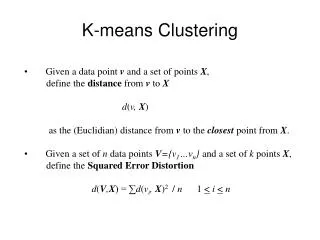
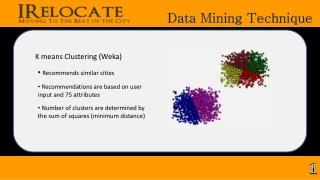
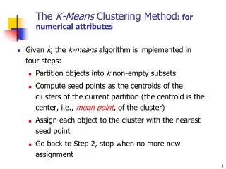
International Journal of Trend in Scientific Research and Development (IJTSRD) ISSN: 2456-6470 traditional methods for measuring tumor volumes are not reliable and are error sensitive. Nursing AND operation. This reflects the means the camera works and the way the info is keep within the pc, however it doesn't correspond to the means that folks acknowledge color. Therefore, the HSL and HSV color models ar a lot of usually used. it's conjointly attainable to use the CMYK color model once applied to a stack of pictures, typical in Medical imaging, the ensuing segmentation are often wont to produce 3D reconstructions with the assistance of interpolation algorithms like walking cubes. In several image process books, the image origin is outlined to be at (xylem)=(0,0).The next coordinate values on the primary row of the image square measure (xylem)=(0,1).It is necessary to stay in mind that the notation (0,1) is employed to suggest the second sample on the primary row. It doesn't mean that these square measure the particular values of physical coordinates once the image was sampled. Following figure shows the coordinate convention. Note that x ranges from zero to M-1 and y from zero to N-1 in number increments. The coordinate convention employed in the tool cabinet to denote arrays is totally different from the preceding paragraph in 2 minor ways that. First, rather than victimization (xylem) the tool cabinet uses the notation (race) to point rows and columns. Note, however, that the order of coordinates is that the same because the order mentioned within the previous paragraph, within the sense that the primary component of a coordinate topples, (alb), refers to a row and also the second to a column. the opposite distinction is that the origin of the arrangement is at (r, c) = (1, 1); thus, r ranges from one to M and c from one to N in whole number increments. IPT documentation refers to the coordinates. Less oft the tool cabinet additionally employs another coordinate convention referred to as abstraction coordinates that uses x to ask columns and y to refers to rows. this can be the other of our use of variables x and y. contours when image The best method of picture segmentation is referred to as the thresholding technique. Several famous strategies are utilized in enterprise which include the most entropy technique, Otsu's approach (maximum variance), and et al. okay-means clustering also can be used. III. CLUSTERING METHODS In statistics and machine learning, the k-means formula could be a agglomeration formula to partition n objects into k clusters, wherever k < n. it's just like the expectation-maximization formula for mixtures of Gaussians therein they each commit to notice the centers of natural clusters within the information. The model needs that the thing attributes correspond to components of a vector area. the target it tries to realize is to reduce total intra-cluster variance, or, the square error operate. The k-means agglomeration was fancied in 1956. the foremost common style of the formula uses associate unvaried refinement heuristic called Lloyd's formula. Lloyd's formula starts by partitioning the input points into k initial sets, either arbitrarily or mistreatment some heuristic information. It then calculates the mean purpose, or center of mass, of every set. It constructs a replacement partition by associating every purpose with the nearest center of mass. II. THRESHOLD SELECTION The key parameter within the thresholding method is that the alternative of the edge price (or values, as mentioned earlier). many completely different strategies for selecting a threshold exist; users will manually opt for a threshold price, or a thresholding formula will cypher a price mechanically, that is thought as automatic thresholding. A additional subtle approach may well be to form a bar graph of the image element intensities and use the vale purpose because the threshold. Then the centroids square measure recalculated for the new clusters, and formula continual by alternate application of those 2 steps till convergence, that is obtained once the points now not switch clusters (or instead centroids aren't any longer changed). Lloyd's formula and k-means square measure typically used synonymously, however in point of fact Lloyd's formula could be a heuristic for resolution the k- means drawback, like sure mixtures of beginning points and centroids, Lloyd's formula will in reality converge to the incorrect answer. alternative variations exist, however Lloyd's formula has Thresholding is termed adaptational thresholding once a special threshold is employed for various regions within the image. this might even be called native or dynamic thresholding. Color pictures also can be threshold. One approach is to designate a separate threshold for every of the RGB elements of the image so mix them with Associate in @ IJTSRD | Available Online @ www.ijtsrd.com | Volume – 2 | Issue – 4 | May-Jun 2018 Page: 724
International Journal of Trend in Scientific Research and Development (IJTSRD) ISSN: 2456-6470 remained well-liked, as a result of it converges very quickly in observe. In terms of performance the formula isn't absolute to come back a worldwide optimum. the standard of the ultimate answer depends mostly on the initial set of clusters, and may, in observe, be abundant poorer than the world optimum. Since the formula is very quick, a typical methodology is to run the formula many times and come back the simplest agglomeration found. A downside of the k-means formula is that the quantity of clusters k is associate input parameter. associate inappropriate alternative of k could yield poor results. The formula conjointly assumes that the variance is associate applicable live of cluster scatter. therefore the region's mean, δ, is employed as a live of similarity. The pel with the littlest distinction measured this manner is allotted to the various region. This method continues till all pixels square measure allotted to a neighborhood. Seeded region growing needs seeds as further input. The segmentation results square measure addicted to the selection of seeds. Unseeded region growing could be a changed rule that does not need specific seeds. It starts off with one region A1 – the pel chosen here doesn't considerably influence final segmentation. If not, then the pel is taken into account considerably totally different from all current regions Ai and a replacement region associate + one is made with this pel. One variant of this method, planned by Haralick and Shapiro (1985), is predicated on pel intensities. The mean and scatter of the region and therefore the intensity of the candidate pel is employed to figure a take a look at datum. Otherwise, the pel is rejected, and is employed to make a replacement region. A special region growing methodology segmentation (see conjointly lambdaconnectedness). it's supported pel intensities and neighborhood linking methods. A degree of property (connectedness) are going to be calculated supported a path that's fashioned by pixels. IV. HISTOGRAM BASED METHODS Histogram-based economical in comparison to alternative image segmentation strategies as a result of they generally need just one submit to the pixels. during this technique of image classification distance metric and integrated region matching area unit acquainted. Histogram-based approaches may be quickly tailored to occur over multiple frames, whereas maintaining their single pass potency. The bar graph are often exhausted multiple fashions once multiple frames area unit thought of. an equivalent approach that's loving one frame are often applied to multiple, and when the results area unit integrated, peaks and valleys that were antecedently tough to spot area unit additional seemingly to be distinguishable. The bar graph may be applied on a per picture element basis wherever the knowledge result area unit wont to verify the foremost frequent color for the picture element location. This approach segments supported active objects and a static surroundings, leading to a unique kind of segmentation helpful in Video trailing. strategies area unit terribly is termed λ-connected VI. SPLIT-AND-MERGE METHODS Split-and-merge segmentation is predicated on a quad-tree partition of a picture. it's generally referred to as quad-tree segmentation. This method continues recursively till no additional splits or merges area unit potential. once a special organisation is concerned within the implementation of the algorithmic program of the strategy, its time complexness will reach O(nlogn), associate degree optimum algorithmic program of the strategy. A.Edge detection VII. Neural networks segmentation To section associate degree object from a picture but, one wants closed region boundaries. Edge is nothing however boundary between 2 pictures. Edge detection technique refers to the identification and locating the sharp discontinuities within the image. Neural Network segmentation depends on process little areas of a picture victimization a man-made neural network[30] or a collection of neural networks. Pulse-coupled neural networks (PCNNs) area unit neural models planned by modeling a cat’s cortical area and developed for superior biomimetic image process. The Eckhorn straightforward and effective tool for finding out little mammal’s cortical area, and was presently recognized as having vital application potential in image process. V. REGION GROWING METHODS model provided a The first region growing methodology was the seeded region growing methodology. This methodology takes a group of seeds as input along side the image. The seeds mark every of the objects to be segmental. The distinction between a pixel's intensity worth and @ IJTSRD | Available Online @ www.ijtsrd.com | Volume – 2 | Issue – 4 | May-Jun 2018 Page: 725
International Journal of Trend in Scientific Research and Development (IJTSRD) ISSN: 2456-6470 and background. These regions is thought of as four completely different categories. Therefore, associate input image has to be divided into these four categories. so as to avoid the probabilities of misclassification, the outer elliptical formed object ought to be removed. By removing this object we are going to get obviate non brain tissues and can be left with solely soft tissues. during this experiment we've used T1, T2 and palladium weighted brain MRIs. These pictures posses same size and same component intensity values. The pixels from the image into consideration is meant to be sorted in anybody of the same category. Finally, by applying sure post process operations, the growth region is extracted. Figure shows the methodology of this work. The process uses K-means algorithmic program for determination clump downside this algorithmic program aims at minimizing associate objective operate, during this case a square error operate. Mathematically, this objective operate is drawn as: VIII.DESIGN ASPECTS A.Region growing method The technique isn't absolutely automatic, i.e. it needs user interaction for the choice of a seed and second the tactic fails in manufacturing acceptable leads to a natural image. It solely works nonuniform areas. Some segmentation ways like "Thresholding" win this goal by yearning for the boundaries between regions supported discontinuities in grey levels or color properties. Region-based segmentation could be a technique for determinant the region directly. the essential formulation for Region-Based Segmentation is: Where may be a chosen distance live between a knowledge purpose xi(j) and also the cluster centrecj, is AN indicator of the space of the n information points from their individual cluster centre’s. Image is browse from information. The image contains the bone tissues. These tissues area unit non brain parts. Therefore, they must be removed within the preprocessing step. The presence of those tissues would possibly cause misclassification. Figure shows a picture of the brain with bone seen as AN outer elliptical ring. In figure this elliptical ring is removed and that we area unit left with solely soft tissues. this is often done by using the subsequent morphological perform, i.e. erosion and dilation. Mathematically, these functions may be expressed as: Mathematically, these functions can beexpressed as: B.Supervised and unsupervised segmentation methods Tried to phase the degree as a full exploitation KNN and each arduous and fuzzy c-means clump. Results showed, however ,that there seems to be enough knowledge nonuniformity between slices to stop satisfactory segmentation. categoryification permits US to own enough notable pixels to come up with representative parameters for every class of interest. In associate unsupervised classification pre hand information of categories isn't needed. It usually workers some clump algorithmic program for classifying a picture knowledge. in keeping with KNN, cc and Parzen window classifiers area unit supervised classification algorithmic program. Whereas, unsupervised algorithmic program includes: KMeans, minimum distance, most distance and stratified clump etc. A brain Image consists of 4 regions i.e. nerve tissue (GM), nerve tissue (WM), cerebrospinalfluid (CSF) Supervised classification C.White matter White matter is one among the 2 elements of the central system and consists principally of myelinate @ IJTSRD | Available Online @ www.ijtsrd.com | Volume – 2 | Issue – 4 | May-Jun 2018 Page: 726
International Journal of Trend in Scientific Research and Development (IJTSRD) ISSN: 2456-6470 daxons. Its white color is thanks to its usual preservation in methanal. A twenty year-old male has around 176,000 kilometer of medullated axons in his brain. The other main part of the brain is gray matter (actually chromatic tan thanks to blood capillaries). a 3rd coloured part found within the brain that seems darker thanks to higher levels of animal pigment in dopaminergic neurons than its near areas is that the substantianigra. Note that nervous tissue will typically seem darker than gray matter on a microscopeslide owing to the sort of stain used. Men have slightly a lot of nervous tissue than females each in volume and long of medullated axons. At the age of twenty, the whole length of medullated fibers in males is 176,000 metric linear unit whereas that of a feminine is 149,000 km. there's a decline in total length with age of regarding 100% every decade specified a person at eighty years mature has ninety seven,200 metric linear unit and a feminine eighty two,000 km. Most of this reduction is because of the loss of dilutant fibers. White matter is that the tissue through that messages pass between completely different areas of nerve tissue at intervals the system. employing a network as AN analogy, the grey matter will be thought of because the actual computers themselves, whereas the nervous tissue represents the network cables connecting the computers along. The nervous tissue is white thanks to the fatty substance (myelin) that surrounds the nerve fibers (axons). This medulla is found in the majority long nerve fibers, ANd acts as an electrical insulation. This is often vital as a result of it permits the messages to pass quickly from place to position. There square measure 3 completely different types of tracts at intervals the white matter: one. Projection tracts, that send action potentials from the cortex to different brain regions, out of the brain to muscles, or into the brain from sense receptors. 2. Commissural tracts, that carry data between the left and right hemispheres of the brain over bridges called commissures. These tracts enable the 2 hemispheres of the brain to speak with each other. 3. Association tracts, that carry data between lobes at intervals an equivalent hemisphere. Long association fibers connect completely different|completely different} lobes of a hemisphere with each other and short association fibers connect different gyri at intervals one lobe. The brain generally (and particularly a child's brain) will adapt to white-matter injury by finding various routes that bypass the broken white-matter areas, and might so maintain sensible connections between the assorted areas of nerve tissue. Unlike nerve tissue, that peaks in development in an exceedingly person's twenties, the nervous tissue continues to develop, and peaks in time of life. This claim has been controversial in recent years, however. A 2009 paper by Gregorian calendar month Scholz and colleagues used diffusion tensor imaging (DTI) to demonstrate changes in nervous tissue volume as a results of learning a brand new motor task (juggling). The study is vital because the initial paper to correlate motor learning with nervous tissue changes. researchers had thought of this sort of learning to be Figure 1: Shows the micro graph showing the white matter {different|totally Figure 2: Shows the Human brain right dissected lateral view Structure White matter consists of bundles of myelinatednerve cell processes (or axons), that connect varied grey substance areas (the locations of somatic cell bodies) of the brain to every alternative, and carry nerve impulses between neurons. The total variety of long vary fibers among a neural structure is a pair of of the full variety of corticocortical fibers and is roughly constant variety as people who communicate between the 2 hemispheres in pathway. Cerebral- and spinal nerve tissue don't contain dendrites, which may solely be found in gray substance beside neural cell bodies, and shorter axons. nerve tissue in nonelderly adults is one.7-3.6% blood. Previously, several @ IJTSRD | Available Online @ www.ijtsrd.com | Volume – 2 | Issue – 4 | May-Jun 2018 Page: 727
International Journal of Trend in Scientific Research and Development (IJTSRD) ISSN: 2456-6470 solely mediate by dendrites, that don't seem to be gift in nervous tissue. The authors counsel that electrical activity in axons might regulate myelination in axons. Similarly, the cause is also gross changes within the diameter or packing density of the axone. White matter forms the majority of the deep elements of the brain and also the superficial elements of the funiculus. Aggregates of nerve tissue like the basal ganglia (caudate nucleus, putamen, globuspallidus, karyon, nucleus accumbens) and brain stem nuclei (red nucleus, substantianigra, nervus nuclei) square measure unfold at intervals the cerebral nervous tissue. The neural structure is structured in an exceedingly similar manner because the neural structure, with a superficial mantle of neural structure cortex, deep neural structure nervous tissue (called the "arbor vitae") and aggregates of nerve tissue encircled by deep neural structure nervous tissue (dentate nucleus, orbicular nucleus, emboliform nucleus, and fastigial nucleus). The fluid-filled cerebral ventricles (lateral ventricles, ventricle, Sylvian aqueduct, fourth ventricle) also are placed deep at intervals the cerebral nervous tissue. Multiple Sclerosis (MS) is one in every of the foremost common diseases that have an effect on nervous tissue. In MS lesions, the medulla defend round the axons has been destroyed by inflammation. Changes in nervous tissue called amyloid plaques square Alzheimers and different neurodegenerative diseases. nervous tissue injuries ("axonal shearing") is also reversible, whereas nerve tissue regeneration is a smaller amount seemingly. different changes that unremarkably occur with age embrace the event of leukoaraiosis, that may be a concentration of the nervous tissue which will be caused by a range of conditions, together with loss of medulla, nerve fibre loss, and a breakdown of the barrier. The study of nervous tissue has been advanced with the neuroimaging technique referred to as diffusion tensor imaging wherever resonance imaging (MRI) brain scanners square measure used. As of 2007, over 700 publications are printed on the topic. colour distinction arises chiefly from the achromatic colour of medulla. In living tissue, grey substance really encompasses a greybrown color, that comes from capillary blood vessels and neuronic cell bodies. Grey matter is created from neuronic cell bodies. The grey substance includes regions of the brain concerned in muscle management, sensory perception like seeing and hearing, memory, emotions, and speech. Grey matter is distributed at the surface of the cerebral hemispheres (cerebral cortex) and of the neural structure (cerebellar cortex), furthermore as within the depths of the neural structure (thalamus; hypothalamus; neural structure, basal ganglia - basal ganglion, globuspallidus, nucleus accumbens; septate nuclei), neural structure (deep neural structure nuclei - nucleus, global nucleus, emboliform nucleus, fastigial nucleus), brain stem (substantianigra, red nucleus, olivary nuclei, nerve nuclei) and spinal grey substance (anterior horn, lateral horn, posterior horn). Significant positive correlations have been found between gray matter volume in elderly persons and measures of semantic and short-term memory. No significant correlations with white matter volume were found. These results suggest that individual variability in specific cognitive functions that are relatively well preserved with aging is accounted for by the variability of gray matter volume in healthy elderly subjects. measure related to Some structural variations in nerve tissue could also be related to psychiatrical disorders. There was no distinction in whole-brain nerve tissue volume between patients with bipolar I disorder and healthy controls. Subjects with bipolar I disorder had smaller volumes within the left inferior membrane bone lobe, right superior convolution, right middle convolution, and left caudate. solely the quantity of the correct middle convolution was correlate with length of ill health and also the variety of episodes in patients. Older smokers lose gray matter and psychological feature operate at a bigger rate than non-smokers. Chronic smokers United Nations agency quit throughout the study lost fewer brain cells and preserved higher intellectual operate than those that continuing to smoke. D.Grey matter Grey matter (or grey matter) could be a major element of the central systema nervosum, consisting of neuronalcell bodies, neuropil (dendrites and each unmyelinatedaxons and myelinated axons), interstitial tissue cells (astroglia and oligodendrocytes) and capillaries. grey substance contains neural cell bodies, in distinction to substantia alba, that doesn't and principally contains myelinated nerve fiber tracts. the @ IJTSRD | Available Online @ www.ijtsrd.com | Volume – 2 | Issue – 4 | May-Jun 2018 Page: 728
International Journal of Trend in Scientific Research and Development (IJTSRD) ISSN: 2456-6470 equation. Infants and patients shunted as babies could have significantly sudden relationships between pressure and bodily cavity size, presumably due partially to blood pressure dynamics. this could have vital treatment implications, however the underlying pathophysiology must be any explored. CSF connections with the systema lymphaticum are incontestable in many class systems. Preliminary information counsel that connections kind round the time that the CSF humour capability of the plexus is developing (in utero). these CSF-lymph Figure 3: Shows the chief ganglionic categories IX. SIMULATION RESULTS A.Cerebrospinal fluid There is also some relationship between CSF disorders, as well as abnormality and impaired CSF humour transport. CSF will be tested for the designation of a range of neurologic diseases.[9] it's sometimes obtained by a procedure known as centesis. Removal of CSF throughout centesis will cause a severe headache when the fluid is removed, as a result of the brain hangs on the vessels and nerve roots, and traction on them stimulates pain fibers. The pain will be mitigated by intrathecal injection of sterile isotonic saline. centesis is performed in a shot to count the cells within the fluid and to sight the amount ofsuper molecule and aldohexose. These parameters alone are also extraordinarily useful within the designation of subarachnoid hemorrhage and central system infections (such as meningitis). Moreover, a CSF culture examination could yield the organism that has caused the infection. By exploitation a lot of refined ways, like the detect ion of the oligoclonal bands, associate degree current inflammatory condition (for example, multiple sclerosis) will be recognized. A beta-2 beta globulin assay is extremely specific and sensitive for the detection for, e.g., CSF discharge. There square measure quantitative distributions of variety of proteins within the CSF. In general, orbicular proteins and simple protein square measure in lower concentration in cavity CSF compared to body part or cisternal fluid. Cerebrospinal fluid (CSF), Liquor cerebrospinal is, is a clean, colorless, salty bodily fluid, that occupies the subarachnoid area and the ventricular device around and in the mind and spinal cord. In essence, the mind "floats" in it. The CSF occupies the space between the arachnoid mater (the center layer of the brain cover, meninges), and the pia mater (the layer of the meninges closest to the brain). It constitutes the content material of all intracerebral (inside the mind, cerebrum) ventricles, cisterns, and sulci (singular sulcus), as well as the important canal of the spinal twine. It acts as a "cushion" or buffer for the cortex, imparting a fundamental immunological protection to the brain in the cranium. mechanical and variations within the CSF serves four primary purposes: Figure 4: Shows a vials of CSF 1. Buoyancy: The actual mass of the human brain is set 1400 grams; but, the internet weight of the brain suspended inside the CSF is equal to a mass of 25 grams. The brain therefore exists in neutral buoyancy, which allows the brain to hold its density without being impaired via its own weight, which would reduce off blood supply and kill neurons in the lower sections without CSF. B.Circulation When CSF pressure is elevated, cerebral blood flow is also constricted. once disorders of CSF flow occur, they'll so have an effect on not solely CSF movement however conjointly craniospinal compliance and also the intracranial blood flow, with subsequent somatic cell and interstitial tissue vulnerabilities. The blood vessel system is additionally necessary during this @ IJTSRD | Available Online @ www.ijtsrd.com | Volume – 2 | Issue – 4 | May-Jun 2018 Page: 729
International Journal of Trend in Scientific Research and Development (IJTSRD) ISSN: 2456-6470 2. Protection: CSF protects the brain tissue from injury when jolted or hit. In certain conditions consisting of car accidents or sports activities injuries, the CSF cannot protect the brain from forced touch with the skull case, inflicting hemorrhaging, brain damage, and from time to time loss of life. parameters alone may be extremely beneficial in the diagnosis of subarachnoid hemorrhage and central nervous system infections (such as meningitis). Moreover, a CSF culture examination may yield the microorganism that has caused the infection. By using more sophisticated methods, such as the detection of the oligoclonal bands, an ongoing inflammatory condition (for example, multiple sclerosis) can be recognized. A beta-2 transferrin assay is highly specific and sensitive for the detection for, e.g., CSF leakage. 3. Chemical stability: CSF flows throughout the inner ventricular system in the brain and is absorbed back into the bloodstream, rinsing the metabolic waste from the central nervous system through the bloodbrain barrier. This allows for homeostatic regulation of the distribution of neuroendocrine factors, to which slight changes can cause problems or damage to the nervous system. for example, excessive glycine concentration disrupts temperature and blood strain manipulate, and excessive CSF pH causes dizziness and syncope. 4. Prevention of brain ischemia: The prevention of brain ischemia is made by lowering the amount of CSF within the restricted space inside the skull. This decreases overall intracranial pressure and allows blood perfusion. While CSF strain is multiplied, cerebral blood waft can be constricted. Whilst disorders of CSF flow occur, they will therefore affect not handiest CSF movement but also craniospinal compliance and the intracranial blood float, with next neuronal and glial vulnerabilities. The venous gadget is likewise vital on this equation. Infants and patients shunted as small kids may have specifically sudden relationships between stress and ventricular size, probably due in part to venous strain dynamics. This can have significant treatment implications, however the underlying pathophysiology needs to be further explored. CSF connections with the lymphatic system have been demonstrated in several mammalian systems. Preliminary data suggest that these CSF- lymph connections form around the time that the CSF secretory capacity of the choroid plexus is developing (in utero). There may be some relationship between CSF disorders, including hydrocephalus and impaired CSF lymphatic transport. CSF can be tested for the diagnosis of a variety of neurological diseases.[9] It is usually obtained by a procedure called lumbar puncture. Removal of CSF during lumbar puncture can cause a severe headache after the fluid is removed, because the brain hangs on the vessels and nerve roots, and traction on them stimulates pain fibers. The pain can be relieved by intrathecal injection of sterile isotonic saline. Lumbar puncture is performed in an attempt to count the cells in the fluid and to detect the levels of protein and glucose. These Figure 5: Shows the methodology @ IJTSRD | Available Online @ www.ijtsrd.com | Volume – 2 | Issue – 4 | May-Jun 2018 Page: 730
International Journal of Trend in Scientific Research and Development (IJTSRD) ISSN: 2456-6470 Figure 6: Shows the image with the outer ring skull Figure 8: Shows the noisy image Figure 9: Shows the enhanced image Figure 7: Shows the removing skull tissues X. CONCLUSION & FUTURE SCOPE To test the algorithmic rule, white Gaussian noise is further to the input image. This image is then processed for improvement. Perona and leader model is employed for this purpose. This modeless partial equation for image de-noising and improvement. The model smoothens the image while not losing vital details with the assistance of following relation. It has been determined that once Perona and leader model is combined with K-means algorithmic rule, it produces reliable results. Attributable to un- supervised nature of the approach, the projected system is economical and is a smaller amount error sensitive. It is deduced from the results that un- supervised segmentation strategies area unit higher than the supervised segmentation strategies. as a result of for victimization supervised methodology plenty of pre-processing is required. Additional significantly, the supervised segmentation methodology needs respectable quantity of coaching and testing information that relatively complicates the method. Whereas, this study is applied to the negligible quantity of information with reliable results. However, it's going to be noted that, the utilization of K-Means bunch methodology is fairly straightforward when put next with oftentimes used segmentation I(x, y, t) is the intensity value of a pixel at sampling position (x, y) and scale t and f(x, y, t) is thediffusivity acting on the system. The diffusivity function in Peronan and Malik mode is given by thefollowing mathematical relation @ IJTSRD | Available Online @ www.ijtsrd.com | Volume – 2 | Issue – 4 | May-Jun 2018 Page: 731
International Journal of Trend in Scientific Research and Development (IJTSRD) ISSN: 2456-6470 fuzzy bunch strategies. potency and providing straightforward output area unit basic options of K- Means bunch methodology. to visualize the accuracy of the projected methodology, commonplace deviations of fresh image, creaky image containing white mathematician noise and increased image. mean and Ananthagiri Vijaya Saradhi, REFERENCES M.Tech Scolar, Digital Image Processing, Nalanda Institute of Engineering and Technology (NIET), Guntur District, Andhra vijayananthagiri@gmail.com 1.M. Mancas, B. Gosselin, B. Macq, 2005, "Segmentation Using GrowingThresholding", Proc. of the Electronic Imaging Conference of the International Society forOptical Imaging (SPIE/EI 2005), San Jose (California, USA). 2.Dong-yong Dai; Condon, B.; Hadley, D.; Rampling, R.; "Intracranialdeformation caused by brain tumors: assessment of 3-D resonanceimaging"IEEE Transactions on Medical Imaging Volume 1993Page(s):693 – 702 3.Dzung L. Pham, ChenyangXu, Jerry L. Prince;"A Survey of Current Methods in MedicalMedical Image Segmentation" Technical Report JHU / ECE 99-01, Department Computer Engineering. The Johns Hopkins University, Baltimore MD21218, 1998. 4.Chowdhury, M.H.; thresholding techniques" RimConference on Communications, Computers, and Signal Processing, 1995. Proceedings.17-19 May 1995 Page(s):585 – 58 5.Norbert Rőhrl, Jose GaliaWeidl, "ComputerAssisted Classification of Brain Tumors", Springer BerlinHeidelberg, pp. 55-60, 2008. 6.Wen, P, Zheng, L and Zhou, J, "Spatial credibilistic clusteringalgorithm in noise image segmentation", IEEE InternationalConference on Industrial Engineering EngineeringManagement, pp: 543 - 547, 2007. Pradesh, India, a Region Teasdale, G.; surface by magnetic 12, Issue 4, Dec. L. Srinivas, Assistant Professor, Digital Image Processing, Nalanda Institute of Engineering and Technology (NIET), Guntur District, Andhra Pradesh, India. ofElectrical and Little, W.D,;"Image IEEE Pacific Iglesias-Rozas and and @ IJTSRD | Available Online @ www.ijtsrd.com | Volume – 2 | Issue – 4 | May-Jun 2018 Page: 732