Download
1 / 17
170 likes | 182 Views
Journal of Molecular Genetics and Medicine publishes novel, peer-reviewed research articles, short communication, case reports, review articles and many more, in broad sections of molecular biology, genetics, chromosomal structure, transcription and translation and its applications in medicine by gene therapy. Molecular medicine is a broad field, where physical, chemical, biological, bioinformatics and medical techniques are used to describe molecular structures and mechanisms, identify fundamental molecular and genetic errors of disease, and to develop molecular interventions to correct them.<br>Manuscripts submitted by the authors will serve as useful resources for future work in the field. Alternatively, manuscripts may describe a novel method of data analysis, which could be applied to publicly available data sets. Manuscripts submitted should provide a new approach to major incurable diseases. The editorial manager system facilitates a user friendly article submission, review and publication. Manuscripts that are thoroughly peer reviewed would ensure the best standards in the industry. The articles will be managed electronically, examined by a scientific committee and anonymous evaluators and published every month in HTML and PDF formats.<br>

E N D
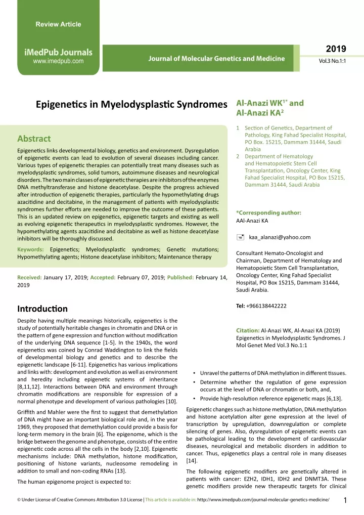
Review Article 2019 iMedPub Journals www.imedpub.com Journal of Molecular Genetics and Medicine Vol.3 No.1:1 Al-Anazi WK1* and Al-Anazi KA2 Epigenetics in Myelodysplastic Syndromes 1 Section of Genetics, Department of Pathology, King Fahad Specialist Hospital, PO Box. 15215, Dammam 31444, Saudi Arabia 2 Department of Hematology and Hematopoietic Stem Cell Transplantation, Oncology Center, King Fahad Specialist Hospital, PO Box 15215, Dammam 31444, Saudi Arabia Abstract Epigeneticslinks developmental biology, genetics and environment. Dysregulation of epigenetic events can lead to evolution of several diseases including cancer. Various types of epigenetic therapies can potentially treat many diseases such as myelodysplastic syndromes, solid tumors, autoimmune diseases and neurological disorders. The two main classes of epigenetic therapies are inhibitors of the enzymes DNA methyltransferase and histone deacetylase. Despite the progress achieved after introduction of epigenetic therapies, particularly the hypomethylating drugs azacitidine and decitabine, in the management of patients with myelodysplastic syndromes further efforts are needed to improve the outcome of these patients. This is an updated review on epigenetics, epigenetic targets and existing as well as evolving epigenetic therapeutics in myelodysplastic syndromes. However, the hypomethylating agents azacitidine and decitabine as well as histone deacetylase inhibitors will be thoroughly discussed. *Corresponding author: AAl-Anazi KA kaa_alanazi@yahoo.com Keywords: Epigenetics; Myelodysplastic syndromes; Genetic mutations; Hypomethylating agents; Histone deacetylase inhibitors; Maintenance therapy Consultant Hemato-Oncologist and Chairman, Department of Hematology and Hematopoietic Stem Cell Transplantation, Oncology Center, King Fahad Specialist Hospital, PO Box 15215, Dammam 31444, Saudi Arabia. Received:January 17, 2019; Accepted: February 07, 2019;Published:February 14, 2019 Tel: +966138442222 Introduction Despite having multiple meanings historically, epigenetics is the study of potentially heritable changes in chromatin and DNA or in the pattern of gene expression and function without modification of the underlying DNA sequence [1-5]. In the 1940s, the word epigenetics was coined by Conrad Waddington to link the fields of developmental biology and genetics and to describe the epigenetic landscape [6-11]. Epigenetics has various implications and links with: development and evolution as well as environment and heredity including epigenetic systems of inheritance [8,11,12]. Interactions between DNA and environment through chromatin modifications are responsible for expression of a normal phenotype and development of various pathologies [10]. Citation:Al-Anazi WK, Al-Anazi KA (2019) Epigenetics in Myelodysplastic Syndromes. J Mol Genet Med Vol.3 No.1:1 • Unravel the patterns of DNA methylation in different tissues. • Determine whether the regulation of gene expression occurs at the level of DNA or chromatin or both, and, • Provide high-resolution reference epigenetic maps [6,13]. Epigenetic changes such as histone methylation, DNA methylation and histone acetylation alter gene expression at the level of transcription by upregulation, downregulation or complete silencing of genes. Also, dysregulation of epigenetic events can be pathological leading to the development of cardiovascular diseases, neurological and metabolic disorders in addition to cancer. Thus, epigenetics plays a central role in many diseases [14]. Griffith and Mahler were the first to suggest that demethylation of DNA might have an important biological role and, in the year 1969, they proposed that demethylation could provide a basis for long-term memory in the brain [6]. The epigenome, which is the bridge between the genome and phenotype, consists of the entire epigenetic code across all the cells in the body [2,10]. Epigenetic mechanisms include: DNA methylation, histone modification, positioning of histone variants, nucleosome remodeling in addition to small and non-coding RNAs [13]. The following epigenetic modifiers are genetically altered in patients with cancer: EZH2, IDH1, IDH2 and DNMT3A. These genetic modifiers provide new therapeutic targets for clinical The human epigenome project is expected to: 1 © Under License of Creative Commons Attribution 3.0 License | This article is available in: http://www.imedpub.com/journal-molecular-genetics-medicine/
2019 ARCHIVOS DE MEDICINA ISSN 1698-9465 Journal of Molecular Genetics and Medicine Vol.3 No.1:1 development [15]. Epigenetic events or modifications are frequently reversible, hence inhibition of epigenetic changes may be a valuable therapeutic potential [14,16,17]. Epigenetic and genetic abnormalities play vital roles in cancer initiation and progression by having frequent mutations [18]. Epigenetic alterations in cancer cells affect virtually all cellular pathways that are associated with tumorigenesis [19]. Epigenetic therapy is intended to reprogram neoplastic cells toward a normal state [18]. Epigenetic drugs can restore defective expression of genes involved in: cell cycle control, apoptosis, cell signaling, tumor cell invasion, metastases, angiogenesis, and immune recognition [19]. and aberrant myeloid differentiation, genetic instability, clonal evolution and increased risk of transformation into secondary AML [36-44]. MDSs manifest as heterogeneous diseases ranging from indolent conditions with considerable life expectancy to aggressive conditions resembling AML. Therefore, risk-adapted treatment strategy is mandatory for MDSs as these diseases have highly variable clinical courses [45]. Pathogenesis, etiology and associations Recent studies in humans and in animal models have provided direct evidence that dysplastic hematopoiesis results from the interaction between: bone marrow (BM) microenvironment, hematopoietic stem cells, and stromal mesenchymal stem cells in the BM niche in patients with MDSs [46-54]. Additionally, epigenetic dysregulation plays an important role in the pathogenesis of MDSs [55]. Etiology and associations of MDSs are shown in Table 2 [40,56-63]. Diseases that can be potentially treated with epigenetic therapies include: • Myelodysplastic syndromes (MDSs); • Other hematologic malignancies (HMs) such as: multiple myeloma (MM), chronic myelomonocytic leukemia (CMML), acute myeloid leukemia (AML), Hodgkin lymphoma and cutaneous T-cell lymphoma; • Metabolic and autoimmune diabetes mellitus, rheumatoid arthritis, systemic lupus erythromatosis, multiple sclerosis, systemic sclerosis and Sjögren's syndrome; • Neurodegenerative and psychological disorders such as: Alzheimer's disease, Parkinson's disease, Huntington's disease and amyotrophic lateral sclerosis; and • Miscellaneous disorders such as psoriasis, cardiovascular disorders and idiopathic pulmonary fibrosis [14-16,18,20-26]. Cytogenetics and molecular genetics Techniques that are used for the detection of cytogenetic abnormalities in MDSs include: • Conventional or metaphase cytogenetics to detect visible chromosomal aberrations; • Fluorescence in situ hybridization (FISH) to detect small and hidden chromosomal aberrations; • Spectral karyotyping to detect unknown and complex chromosomal abnormalities; • Single nucleotide polymorphism array (SNP-A) to detect cryptic and complex chromosomal aberrations; • Microarray-based comparative genome hybridization (CGH) to detect uniparental disomy and copy number variation (CNV); • Sequencing-based technologies such as next generation sequencing (NGS) to detect CNV and structural variants as well as unknown mutations and aberrations; and • PCR [64,65]. Conventional cytogenetics and FISH can detect abnormalities in chromosomes: 5, 7 and 8 while array-CGH and PCR can detect the following somatic mutations: ASXL1, EZH2, TP53, TET2, RUNX1, SF3B1 and DNMT3A [65]. disorders such as: There are several classes of epigenetic drugs. The main types of epigenetic therapies and examples of some types are included in Table 1 [14,15,18,22,27-33]. Epigenetic therapy is a novel therapeutic approach that modulates gene expression by targeting the: DNA methylation machinery, histone covalent modification and micro-RNAs (miRNAs) [20]. A major limitation of epigenetic therapy is the lack of specificity and the consequent global induction of epigenetic changes [20]. Treatment with epigenetic agents can reduce chemotherapy resistance in patients with HMs and solid tumors so epigenetic drugs can be added to cytotoxic chemotherapy or targeted therapy in order to derive chemosentitization benefits [34,35]. Methods that are used in the detection of methylation status of gene promoters and the association between methylation status and clinical parameters in patients with HMs include: methylation-specific polymerase chain reaction (PCR), methylation-specific restriction enzyme digestion, Hpall tiny fragment enrichment by ligation-mediated PCR, bisulphite sequencing and pyrosequencing [15]. Cytogenetic abnormalities, gene mutations and recurrent somatic mutations in MDSs are shown in Table 3 [42,62,66,67], Table 4 [45,68,69] and Table 5 [40,45,62,68,70-73] respectively. MDSs are characterized by mutations in more than 40 genes, a complex structure of gene-gene interactions and extensive subclonal diversification [71,73]. The most frequently mutated genes in MDSs are: TET2, SF3B1, ASXL1, DNMT3A, SRSF2, U2AF1, RUNX1, TP53, EZH2, ZRSR2, STAG2, CBL, NRAS, JAK2, SETBP1, IDH1, IDH2 and ETV6 [73-79]. The following mutated genes are considered epigenetic regulators: TET2, IDH1, IDH2, DNMT3A, ASXL1 and EZH2 [79]. Gene mutations that are independently associated with shorter survival and unfavorable outcome include: ASXL1, U2AF1, TP53, SRSF2, CBL, IDH2, SETBP1, DNMT3A, RUNX1 and EZH2 [75,76,80,81]. However, SF3B1 gene mutation has been associated with longer survival and favorable outcome [80,82]. Identification of somatic mutations in patients with MDSs suggests MDSs Introduction to MDSs MDSs comprise a group of biologically and clinically heterogeneous clonal hematopoietic neoplasms characterized by: peripheral cytopenias, dysplastic changes in at least one hematopoietic lineage, ineffective hematopoiesis due to excessive apoptosis 2 This article is available in: http://www.imedpub.com/journal-molecular-genetics-medicine/
2019 ARCHIVOS DE MEDICINA ISSN 1698-9465 Journal of Molecular Genetics and Medicine Vol.3 No.1:1 Table 1 Types and examples of epigenetic drugs. Classes or types of epigenetic drugs DNA methyltransferase inhibitors [DNMTIs] or demethylating agents Histone deacetylase inhibitors [HDACIs] (5 classes: I, IIa, IIb, III and IV) Histone acetylase/acetyltransferase inhibitors Histone demethylase inhibitors Protein methyltransferase inhibitors Sirtuin inhibitors and modulators Bromodomain inhibitors Examples Azacitidine, decitabine, zebularine, S-110 and SGI-1027 Vorinostat, panobinostat, entinostat, givinostat, pracinostat, belinostat, valproic acid, romidespsin, pivanex, CI-994 and ACY-1215 6 thioguanine; fazarabine; pseudoisocitidine; 5 fluoro-2-deoxycitidine and 5,6 dihydro-5-azacitidine Nucleosidic DNA methyltransferase inhibitors Antisense oligonucleotide inhibitors of DNMTs Inhibitors of protein binding to acetylated histone Inhibitors of protein binding to methylated histone Procainamide, hydrallazine, methotrexate, thalidomide, statins, neuroleptics, B-blockers, Fluoroquinolones, isotretinoin, cox-2 inhibitors, synthetic estrogens and general anesthetics. Miscellaneous drugs that have epigenetic activities Table 2 Etiology and associations of myelodysplastic syndromes. 1. Unknown etiology. 2. Old age; more than 50 years. 3. Male gender. 4. Obesity. 5. Tobacco use. 6. Alcohol intake. 7. Sweet syndrome; neutrophilic dermatosis. 8. Vitamin deficiencies: - Folic acid - Vitamin-B12. 9. Infections: - Human immunodeficiency virus. - Tuberculosis. - Brucellosis. 10. Occupational and environmental exposure: solvents, benzene, lead, arsenic, pesticides, herbicides, hair dyes, and agricultural chemicals. 11. Autoimmune disorders: - Systemic lupus erythromatosis. - Fibrosing alveolitis. - Behcet syndrome. - Other vasculitis disorders and seronegative polyarthropathies 12. Therapy-related myelodysplastic syndromes: - Alkylating agents. - Topoisomerase II inhibitors. - Radiotherapy. 13. Bone marrow failure syndromes: - Aplastic anemia. - Diamond Blackfan syndrome. - Fanconi anemia. - Paroxysmal nocturnal hemoglobinuria. - Dyskeratosis congenita. - Congenital neutropenias. 14. Genetic, familial and hereditary disorders: - Ataxia telangiectasia - Down's syndrome - Xeroderma pigmentosa - Trisomy 8 mosaicism - Bloom's syndrome - Neurofibromatosis new targets for therapeutic interventions [68]. For example TP53 mutations, which are less likely to respond to single agent lenalidomide, have been reported to occur in approximately 20% of patients with del 5q, low and intermediate I MDSs [45]. [70,83-87]. After obtaining PB-CF-DNA from plasma or serum, high resolution SNP-A is used for karyotyping then mutation analysis of genes is performed using PCR or sequencing (Sanger, parallel or targeted NGS) [83,85,87]. Studies have shown high concordance rates reaching 100% in cytogenetic or mutational profiles between PB and BM in patients with MDSs [83,86]. Mutations in the following genes can be determined by PB-CF- DNA: SF3B1, DNMT3A, ASXL1, SRSF2, IDH1, IDH2, TET2, U2AF1, ZRSR2, RUNX1, ETV6, NRAS, KRAS, TP53, CBL, JAK2, MPL, CEBPα, Diagnosis of MDSs using peripheral blood Recently, several studies have shown that peripheral blood cell- free DNA (PB-CF-DNA) is safer, easier and even more sensitive for genetic and epigenetic analyses than whole BM samples 3 © Under License of Creative Commons Attribution 3.0 License
2019 ARCHIVOS DE MEDICINA ISSN 1698-9465 Journal of Molecular Genetics and Medicine Vol.3 No.1:1 Table 3 Cytogenetic abnormalities in myelodysplastic syndromes. Risk Category Very Good Good Examples del 11q; -Y Normal cytogenetics; del 20q; del 5q; single or double; del 12p +8; del 7q; i17q; +19; +21; any other single or double abnormality; independent clones -7; inv 3; del3q/t3q; 2 abnormalities including -7/del 7q; complex cytogenetics (3 abnormalities) Complex cytogenetics: >3 abnormalities Intermediate Poor Very Poor Table 4 Genetic mutations in myelodysplastic syndromes (MDSs). Mutated Genes Frequency (%) 15-30 (up to 80% in MDS-RARS) 2-12 5-12 5 5-22 15-26 4-11 10-20 3-7 5-10 5-10 5-6 3 5-10 Prognosis SF3B1 Good, favorable outcome with longer event free survival SRSF2 Poor with short overall survival Poor with rapid transformation into acute myeloid leukemia Neutral Poor Neutral with no impact on survival Mixed evidence Poor Poor Poor Poor Poor Poor Poor U2AF1/U2AF35 ZRSR2 DNMT3A TET2 IDH1/IDH2 ASXL1 EZH2 RUNX1 TP53 BCOR ETV6 NRAS/KRAS - RARS: Refractory anaemia ring sideroblasts Table 5 Recurrent somatic mutations in myelodysplastic syndromes. Pathway DNA methylation (epigenetic regulatory genes) Examples of genetic mutations - DNMT3A - IDH1 - TET 2 - IDH2 - ATM - DLRE1C - FANCL - BRCC3 - TP53 - ASXL1 - EZH2 - NRAS / KRAS - CBL/NF1 - JAK2 - PTPN11 - FLT3 - STAG2 - RAD21 - SMC1A/SMC3 - CTCF - SF3B1 - U2AF1 - SRSF2 - ZRSR2 CEBPA - RUNX1 - BCOR1/ BCORL1 - GATA2 - ETV6/EVI1 DNA repair Chromatin modification Signal transduction (Kinases/RAS pathway) Cohesion complex RNA splicing (splicing factor genes) Transcription factors and transcriptional regulation • Detection of cytogenetic abnormalities and genetic mutations that predict evolution of new clones and disease progression, SETBP1, FLT3, BRAF and NPM1 [70,85-87]. PB-CF-DNA analysis has the following advantages: 4 This article is available in: http://www.imedpub.com/journal-molecular-genetics-medicine/
2019 ARCHIVOS DE MEDICINA ISSN 1698-9465 Journal of Molecular Genetics and Medicine Vol.3 No.1:1 Table 6 The revised international prognostic scoring system (R-IPSS) for myelodysplastic syndromes. Points 1.5 - - < 8 Prognostic variable 0 0.5 - - - 1 2 3 4 Cytogenetics Bone marrow blasts % Hemoglobin (g/dL) Platelet count Very good ≤ 2 ≥ 10 Good >2 - 5% 8 - <10 Intermediate 5% - 10% - Poor > 10 % - Very poor - - ≥ 100 50 - 100 <50 - - - - × 109/L Absolute neutrophil count ≥ 0.8 < 0.8 - - - - - × 109/L Table 7 Current clinical picture of personalized medicine in MDSs. Variable Grading Good Poor Low High High Good risk Poor risk Del 5q Potential clinical consequences Standard treatment including allogeneic HSCT Supportive care only Treatment with erythropoietin stimulating agents No treatment with erythropoietin stimulating agent in case of anemia Treatment with iron chelation Supportive care only Hypomethylating agents and allogeneic HSCT Treatment with lenalidomide Supportive care only Performance status Erythropoietin level Ferritin level Prognostic scoring index (IPSS/R-IPSS) Cytogenetics Good risk Genetic mutations Standard treatment including allogeneic HSCT Intensified surveillance or early pre-emptive therapy in otherwise good-risk MDSs (e.g., by R-IPSS) Poor risk ● MDSs: myelodysplastic syndromes ● HSCT: hematopoietic stem cell transplantation ● IPSS: international prognostic scoring index ● R-IPSS: revised international prognostic scoring index • Establishing the diagnosis of MDSs in patients with cytopenias, • Obviating the need for repeated BM examinations particularly in elderly patients and those with hypocellular or fibrotic BMs, and • Monitoring response to cytotoxic chemotherapy and targeted agents including epigenetic therapies [83,86,87]. risk MDSs with <10% blasts include: • Growth factors such as erythropoietin and granulocyte- colony stimulating factor (G-CSF), • Immune therapies including: corticosteroids, cyclosporine-A and antithymocyte globulin, • Lenalidomide for 5q31, • Decitabine and azacitidine, • Iron chelation and blood transfusion, • Imatinib for t5,12 and 5q33 variant with platelet-derived growth factor receptor (PDGFR)-β; and • Investigational therapies such as clofarabine and homoharringtonine. • For higher-risk (HR) MDSs with ≥ 10% blasts and chromosome 7 abnormalities or complex cytogenetics, therapeutic options include: • Decitabine and azacytidine, • Intensive chemotherapy for younger patients and those with diploid karyotype, • Allogeneic hematopoietic stem cell transplantation (HSCT), • Imatinib for t5,12 and 5q33 variant with PDGFR-β, • Iron chelation and blood transfusion; and • Investigational therapies [37,39,40,42,56,91]. Epigenetic modifying agents that are used in patients with MDSs include: New techniques in the diagnosis of MDSs The following are new techniques that are helpful in establishing the diagnosis of MDSs: • Immunophenotyping by flowcytometry of PB neutrophils and monocytes particularly in low-risk MDSs, • Proliferation index of specific compartments of BM cells that reflects the rate of production of hematopoietic cells in MDSs, and • Measurement of telomere length in PB leukocytes as shorter telomeres have been found to be associated with occupational exposure to paints and pesticides [88-90]. Therapeutic options in MDSs Treatment of MDSs is selected based on: risk stratification by the international prognostic scoring index (IPSS) and the revised IPSS, transfusion needs, percentage of BM blasts and cytogenetic as well as mutational profiles [42]. The revised IPSS in patients with MDSs is shown in Table 6 [38-40]. Therapeutic options for low- 5 © Under License of Creative Commons Attribution 3.0 License
2019 ARCHIVOS DE MEDICINA ISSN 1698-9465 Journal of Molecular Genetics and Medicine Vol.3 No.1:1 Table 8 Drugs in clinical trials and future therapies for myelodysplastic syndromes. Genetic mutations, pathway or target IDH1/IDH2/R132 SF3B1, SRSF2, U2AF1, ZRSR2 TET 2 TP53 including 5q- syndrome Epidermal growth factor receptor Dual inhibitor [phosphoinositide-3 kinase and polo-like kinase] Programmed cell death 1 protein (PD-1) and PD-1 ligand-1(PD- 1L-1) Bone marrow megakaryocytes Drugs in clinical trials FT-2102, AG-881, ivosidenib, venetoclax and enasidenib H3B-8800 (oral) Ascorbic acid (oral and intravenous) and hypomethylating agents APR-246 (intravenous) and decitabine Erlotinib Rigosertib PD-1 and PD-1L1 inhibitors Eltrombopag and romiplostim • Demethylating agents such as: azacitidine licensed for Demethylating agents such as: azacitidine licensed for MDSs and AML, decitabine licensed for HR-MDSs and AML, and zebularine which is still investigational, and • HDACIs which are still investigational and they include: panobinostat, vorinostat, entinostat, belinostat and romidepsin and valpoic acid [69]. 2004, then 5 years later it was granted extended approval for use in HR-MDSs [94,97-99]. Studies have shown that the following groups of patients with MDSs appear to have particular benefit: • Chromosome 7 abnormalities including monosomy 7, • Trisomy 8, • Diploid karyotype, and Prognosis in MDSs • HR-del 5q harboring TP53 mutations [94,100]. Azacitidine is indicated not only in MDSs but also in AML and CMML [94,97-99,101-107]. In patients with MDSs, prognosis is determined by: • IPSS and R-IPSS, • Age, • Performance status, • Comorbid medical conditions, • Transfusion dependence, and • Molecular biomarkers such as somatic mutations that can be detected by several methods including DNA sequencing [92]. Azacitidine is a disease modifying agent that has changed the history of MDSs and it has been shown to impact positively all the 3 cell lines [94,97]. At high doses, azacitidine is cytotoxic and its cytotoxicity results from incorporation into DNA and RNA, while at lower doses the drug has hypomethylating effects as it induces differentiation and demethylation resulting in restoration of normal growth control and differentiation into hematopoietic cells [94,97,99]. The effectiveness of azacitidine was first demonstrated in the following 3 studies: • A single randomized controlled trial comparing azacitidine administered subcutaneously (SC) with best supportive care (observational group) which showed 16% response rate in the study group and 0.0% response rate in the observational group, and • Two single arm studies, in one azacitidine was administered intravenously (IV) and in the other it was given SC and these 2 studies showed response rates of 13% and 19% respectively and these responses were sustained for 11 and 17 months respectively [97,99,102]. Epigenetic Therapies in MDSs HypomethylatingAgents (HMAs) Epigenetic mechanisms such as abnormal DNA methylation are considered the first markers of tumorigenesis [93]. Methylation of the tumor suppressor gene CDKN2B is frequent in patients with MDSs and is usually acquired during disease progression [94]. DNA hypermethylation is well documented in the pathogenesis of MDSs [95,96]. Reversal of unfavorable methylation status in malignant cells has been a subject for epigenetic therapy of cancer using HMAs [93]. Reactivation by demethylation may halt disease progression [94]. Restoration of transcriptionally silenced genes by means of DNA methyltransferase inhibitors (DNMTIs) plays an important role in the current management of MDSs [93]. Thus, methylation status may serve a marker to monitor response to epigenetic therapies [96]. Several studies have shown that azacitidine can: • Prolong survival, • Prolong time to leukemic transformation from 12 to 21 months, • Reduce transfusion requirements of blood products, and Azacitidine: Azacitidine is a pyrimidine nucleoside analog that was chemically synthesized and characterized by Frantisek Sorm et al in Czechoslovakia in the 1960s. It differs from cytosine primarily by the presence of nitrogen at position 5 [97]. Azacitidine is a DNMTI that leads to reduction of DNA methylation in patients with MDSs [93]. Azacitidine was the first HMA to be approved by the food and drug authority in the United States of America (USA-FDA) for the treatment of all subtypes of MDSs in the year • Improve quality of life, while maintaining a relatively safe toxicity profile even in elderly individuals [95,97,101,103- 105,108-112]. Complete remissions (CRs) can be encountered in up to 25% of patients, but unfortunately some patients do not respond to azacitidine possibly due to having inadequate plasma levels of the drug [94]. In order to improve response rates further, azacitidine 6 This article is available in: http://www.imedpub.com/journal-molecular-genetics-medicine/
2019 ARCHIVOS DE MEDICINA ISSN 1698-9465 Journal of Molecular Genetics and Medicine Vol.3 No.1:1 predict response, longer overall survival (OS) and longer duration of response: red blood cell transfusion requirements, performance status, circulating blast cells, doubling of platelet count after first cycle of therapy, type of induction therapy given prior to HSCT, karyotype particularly HR cytogenetics, preceding 5q- syndrome, therapy-related MDSs, and mutational profile particularly TET2, SRSF2, TP53 and KDM6A mutations [45,114,119,120]. However, negative outcome, shorter OS and shorter duration of response to azacitidine have been reported with CDKN2A mutations [121- 125]. Despite the presence of several publications indicating the presence of predictive biomarkers for response to azacitidine, a study that included 128 patients with MDSs and AML treated with azacitidine has shown no clear biomarkers for response to azacitidine and survival that could be identified [121-123]. can be combined with lenalidomide, histone deacetylase inhibitors (HDACIs) and growth factors [97,100]. Although prolonged use of the drug is generally practiced, patients may benefit from a limited number of cycles of azacitidine [113]. The drug can induce complete and partial responses in approximately 50% of patients, these responses are usually not durable or sustainable as most responding patients lose their responses within 2 years [106,113,114]. Azacitidine can be given IV or SC. The standard and approved dose of 75 mg/m2/day for 7 days every 28 days has been proven to show objective response rates, while the other dose schedule of 100 mg/m2/day for 5 days has not been approved although this schedule is given taking into consideration convenience and logistic factors [97-99,102,109,110]. Azacitidine is rapidly absorbed after SC administration and maximum plasma concentration is reached within 30 minutes of SC administration and 10 minutes of IV administration [97,99]. The drug is widely distributed in tissues. Its bioavailability after SC administration is 89% of that after IV administration and plasma half-life is approximately 41 minutes after SC administration and about 22 minutes after IV administration [97,99]. In patients with MDSs treated with azacitidine, P53 expression which is a surrogate for the presence of TP53 mutation does not have negative impact on treatment response indicating that response to azacitidine is independent of P53 expression in patients with HR-MDSs [126]. Thus, the combination of azacitidine and lenalidomide may be beneficial in patients with del 5q harboring TP53 mutations [126]. The adverse effects of azacitidine include: • Gastrointestinal tract (GIT) manifestations such as: nausea, vomiting, diarrhea, and constipation; • Myelosuppression: neutropenia causing febrile neutropenia and infections in addition to thrombocytopenia causing petechiae, ecchymoses and other bleeding complications; • Injection site reactions; • Fever and rigors; • Headache, dizziness and arthralgia; • Liver dysfunction; • Renal failure particularly in patients with hypotension and sepsis; and • Treatment-related mortality (TRM) [94,98,99,101,113-115]. Decitabine Decitabine, 2-deoxy-5-azacitidine, is similar to azacitidine in structure and inhibition of the enzyme DNMT, but has different mechanisms of action [73,127-129]. Decitabine, a cytosine analog, is cytotoxic at high doses and has DNA demethylating activity at low doses [130]. In the year 2006, decitabine was approved by the USA-FDA for the treatment of de novo, secondary and therapy-related MDSs [127]. Although the antitumor activity of decitabine is not fully understood, it may involve one or more of the following: • Reversal of cancer-associated hypermethylation events, • Reactivation of genes that are responsible for cellular differentiation, • Stimulation or induction of immune responses, • Induction of DNA-damage response pathways, • Augmentation of stem cell renewal, and • Changes in the rate of apoptosis or apoptotic response pathways [127]. Oral azacitidine: In early phase clinical trials, oral azacitidine (CC- 486) has been shown to be biologically and clinically active in patients with MDSs. Hence, it is currently evaluated in ongoing phase III clinical trials [108,115]. Oral azacitidine improves convenience and eliminates injection-site reactions and it enables testing of novel longer term low-dose schedules that enhance therapeutic activity of the drug by increasing exposure to circulating malignant cells [108]. Decitabine has been used in the treatment of HR-MDSs, CMML, and AML particularly in elderly individuals [127,128,131-134]. It can be given: in upfront setting in the treatment of MDSs patients, in the maintenance therapy after allogeneic HSCT, and with fludarabine and total body irradiation (TBI) conditioning therapy prior to HSCT [127-129,132,133,135]. In patients with MDSs, decitabine has been used in combination with the following medications: • Traditional Chinese medicine, • Fludarabine and TBI conditioning therapy, • Aclarubicin hydrochloride, cytosine arabinoside and G-CSF, • Low dose chemotherapy particularly cytosine arabinoside, • Tosedostat, and • Valproic acid. Oral azacitidine can be given at doses ranging from 300 mg to 400 mg per day for 14-21 days each cycle [108,115,116]. In patients with MDSs (including lower-risk groups and patients with pre- treatment thrombocytopenia), AML and CMML, oral azacitidine in extended dosing regimens has been shown to be associated with significant DNA hypomethylation effect and overall response rates (ORRs) ranging from 35% to 73% [108,115-118]. The adverse effects of oral azacitidine include: GIT disturbances, myelosuppression, bleeding and TRM [108,115,116]. Response to azacitidine: In patients with MDSs treated with azacitidine, several studies have shown that the following factors 7 © Under License of Creative Commons Attribution 3.0 License
2019 ARCHIVOS DE MEDICINA ISSN 1698-9465 Journal of Molecular Genetics and Medicine Vol.3 No.1:1 • Interference with or inhibition of chaperone protein functions, • Upregulation of endogenous inhibitors of cell cycle progression such as p21 and disruption of cell cycle checkpoints thus causing cell cycle arrest, • Generation of free radicals and induction of autophagy, • Promotion of apoptosis by inhibition of anti-apoptotic proteins, and • Inhibition of angiogenesis and proteasome function [142,143]. However, in patients with MDSs and AML, the combinations of decitabine with these medications have yielded variable responses [55,73,129,131,132,134,136-139]. The side effects of decitabine include: hematologic toxicity with neutropenia causing febrile neutropenia and infections such as pneumonia and thrombocytopenia causing bleeding from various sites; GIT toxicity such as nausea, vomiting, diarrhea and mucositis; hyperbilirubinemia; cardiovascular toxicity; renal failure; fatigue; and hair loss [127,128,133-135]. Several studies and 2 meta-analyses have shown superiority of azacitidine to decitabine in the treatment of patients with MDSs [127]. Hence, the use of decitabine in the treatment of HR-MDSs is not recommended after failure of azacitidine due to short duration of response and poor OS [136,137]. Also, the addition of valproic acid to decitabine has not been associated with improved outcome in patients with MDSs [131]. Unfortunately, resistance to HDACIs frequently evolves and the following mechanisms of resistance have been described: • Increased expression of multidrug resistance-associated proteins; • Enhanced expression of p21 cell cycle protein; • Increased expression of thioredoxin; • Enhanced expression of anti-apoptotic proteins and inability to upregulate pro-apoptotic proteins; • Alterations of HDAC protein levels; • Increased protein signaling via the following pathways: mitogen-activated protein 3-kinase, as well as signal transducer and activator of transcription; and • Activation of nuclear factor kappa light chain enhancer signaling pathway and acetylation of p65 [140]. Clinical activity of HDACIs Disappearance of TP53 mutation has been shown to be an indication of response to decitabine in patients with MDSs and AML [128]. Recovery of platelet count by the second cycle of decitabine therapy can be used as an early predictor marker of improved survival and an increased response rate [138]. Several studies have sought to identify biomarkers that may predict response to decitabine such as: DNA methylation changes, expression of miR-29b, and specific genetic mutations such as: DNMT3A, IDH1, IDH2 and TET2 [128]. However, controversy still exists regarding the predictive value of these mutations. Additionally, none of the above suggested biomarkers is currently used to guide decitabine treatment for individual patients [128]. kinase, phosphoinositide In general, when used as single agents, HDACIs have shown only modest clinical activity in the treatment of patients with MDSs. However, marked responses have been observed in selected subsets of patients and once HDACIs are used in combination with other agents particularly HMAs [142,144]. Nevertheless, a recently published systematic review and a meta-analysis that included 7 clinical studies comprising 922 patients; 458 patients treated with HMAs alone and 464 patients treated with combination of HMAs and HDACIs; showed no significant differences in CR rates, hematologic improvement, ORRs, OS and toxicities between patients treated with HMAs alone or combined therapy [138]. Additionally, while significant results have been achieved with the use of HDACIs in the treatment of lymphomas and MM, efficacy in patients with myeloid malignancies has remained limited [145]. HDACIs in MDSs In the nucleus, DNA is wound around 4 core histone proteins to form nucleosomes that, when compacted, form the condensed structure of chromatin [140]. Histones can be modified by several processes that include: acetylation, methylation, phosphorylation, sumoylation, ubiquitination, and citrullination [140]. Modifications of DNA or histones via methylation or acetylation lead to gene silencing and altered physiology relevant to MDSs [141]. Acetylation, which is one of the main histone modifications associated with gene expression, is controlled by 2 groups of enzymes: histone acetyltransferases and histone deacetylases (HDACs) [140,142]. Classes, mechanisms of action and resistance to HDACIs Obviously, many issues related to HDACIs remain incompletely understood and pose clinical and translational challenges [141]. Hopefully, the recent advances in disease biology and the design of more specific third generation HDACIs may drive the future clinical development of HDACIs in patients with myeloid malignancies in particular [146]. HDACIs are epigenetic agents that act by modifying gene expression to restore the normal differentiation or death programs of transformed cells [143]. They regulate the acetylation of histones as well as non-histone protein targets [142]. There are 5 classes of HDACIs: class I, class IIA, class IIB, class III and class IV [140]. HDACIs have various mechanisms of action that include: • Chromatin remodeling thus permitting re-expression of tumor suppressor genes that are abnormally suppressed or silenced in cancer cells, • Relaxation of DNA, induction of DNA damage and inhibition of DNA repair, Specific HDACIs Vorinostat: Vorinostat, suberoyalanilide hydroxamic acid, is a HDACI that was approved by the US-FDA in December 2006 for the treatment of relapsed or refractory cutaneous T-cell lymphoma [147-149]. It promotes protein acetylation; modulates gene expression; and induces differentiation, growth arrest and apoptosis of tumor cells [148,149]. It has shown promising 8 This article is available in: http://www.imedpub.com/journal-molecular-genetics-medicine/
2019 ARCHIVOS DE MEDICINA ISSN 1698-9465 Journal of Molecular Genetics and Medicine Vol.3 No.1:1 clinical activity against certain hematologic malignancies and solid tumors such as: MDSs, AML, CMML, MM, cutaneous T-cell lymphoma, diffuse large B-cell lymphoma, Hodgkin's lymphoma, in additions to carcinomas of the: breast, prostate, colon and lung [147-154]. myelosuppression with subsequent bleeding and infectious complications, constitutional symptoms, and metabolic as well as electrolytic disturbances [164]. Valproic acid (phenylbutyrate): Valproic acid (VPA), which has been known as an epileptic agent for many years, is a short- chain fatty acid and a HDACI that can reduce proliferation and induce differentiation of myeloid blast cells in patients with MDSs and AML particularly when given in combination with all-trans retinoic acid (ATRA) [166-169]. In patients with MDSs and AML, VPA as a single agent has shown limited clinical activity, but once used in combination with other drugs; such as: azacitidine, ATRA, bortezomib, hydralazine, decitabine or cytotoxic chemotherapeutic agents; it has been reported to be clinically active due to synergistic anti-leukemic activity and positive effects on blood indices with a significant increase in platelet count in particular [131,166-175]. Although VPA is generally well tolerated with modest side effects, moderate to severe hematologic toxicity including myelosuppression and evolution of myelodysplasia has been reported with the prolonged use of the drug [166,176- 182]. The adverse effects are more pronounced once the drug is given in combination with other agents and these include various degrees of myelosuppression, evolution of MDSs and neurotoxicity [131,168,169]. In patients with MDSs, CMML and AML, the efficacy of vorinostat as a single agent is limited but several phase I and II clinical trials using combinations of vorinostat with conventional chemotherapeutic agents such as idarubicin and cytarabine or investigational drugs such as HMAs or lenalidomide have shown more promising results [147,148,150,154]. However, in patients with MDSs and AML, the use of vorinostat in combination with bortezomib or alvocidib has not shown any objective clinical responses [151,153]. As a single agent or in various drug combinations, vorinostat has acceptable toxicity profile with mainly gastrointestinal and constitutional side effects [148,154]. The main adverse effects of the drug include: nausea, vomiting, diarrhea, dehydration, anorexia, fatigue, cytopenias including thrombocytopenia, prolongation of QT interval on electrocardiogram, abnormal liver profile and metabolic disturbances including hypokalemia, hyperglycemia and hypophosphatemia [147,148,150-154]. Panobinostat: Panibinostat is a potent oral pan-deacetylase inhibitor of HDAC enzymes implicated in cancer development and progression that has been approved for the treatment of MM in the USA, Japan and Europe [155,156]. It modulates the acetylation of histone proteins and protein chaperones in malignant cells and its epigenetic regulation is primarily modulated through inhibition of class I histone deacetylase enzymes leading to: increased histone acetylation, relaxation of chromatin and alteration of expression of certain genes including tumor suppressor genes [155]. Other HDACIs: Unfortunately, clinical trials combining entinostat or pracinostat with azacitidine in the treatment of patients with MDSs and AML have not shown any additional beneficial effects [183,184]. LBH589 is a novel HDACI that inhibits proliferation and induces apoptosis in tumor cell lines [185]. A phase I study on the IV use of LBH589 in a limited number of patients with HMs including AML and MDSs has shown consistent but transient anti- leukemic and biological effects [185]. A phase II clinical trial on the use of belinostat in the treatment of 21 patients with MDSs showed only 5% ORR with significant grade 3-4 toxicities so the study was ultimately terminated [186]. Phase I and II clinical trials on the use of panobinostat in low or intermediate risk MDSs have shown either limited or no meaningful clinical activity [157,158]. However, the use of the drug in combination with azacitidine in the treatment of MDSs, CMML and AML has shown variable clinical activity [159-161]. Additionally, the use of panobinostat in the maintenance therapy after allogeneic HSCT in patients with HR-MDSs and AML has been shown to prolong OS and to reduce rate of relapse of the primary disease [162]. The following adverse effects have been reported with the use of panobinostat: constitutional symptoms, GIT upset, BM suppression, infections, neuropathy and metabolic disturbances [155,159-162]. Challenges in the Current Management of MDSs Thrombocytopenia and its future therapies in MDSs Thrombocytopenia, which is commonly encountered in patients with MDSs, has multifactorial etiology and its associated bleeding complications represent a major cause of morbidity and mortality [187-190]. The thrombopoietin agonists, eltrombopag and romiplostim, have shown clinical activity in trials performed in patients with MDSs and thus they represent a potential alternative therapeutic option to platelet transfusions [187,191]. Several phase I and phase II clinical trials have shown not only safety but also efficacy of eltrombopag in the treatment of thrombocytopenia in patients with advanced MDSs [190,192- 194]. Eltrombopag has been shown to increase megakaryocytic differentiation thus leading to the formation of normal megakaryocytic clones [195]. A single phase II clinical trial on the use of romiplostim in patients with low and intermediate-risk MDSs receiving azacitidine therapy has shown clinical benefit [189]. More prospective and ramdomized clinical trials on the Romidepsin: Romidepsin (depsipeptide) is a bicyclic peptide that showed class I selective HDACI activity in the year 1998. Subsequently, it was approved by the USA-FDA for the treatment of cutaneous T-cell lymphoma [163,164]. Promising results have emerged from early clinical trials supporting the use of romidepsin in conjunction with other drugs for the treatment of other types of lymphoma, MM as well as certain solid tumors [163]. As monotherapy in unselected patients with MDSs and AML, romidepsin has shown limited clinical activity [164]. However, its use in patients with core binding factor AML has shown differential anti-leukemic and molecular activity [165]. The adverse effects of romidepsin include: GIT toxicity, 9 © Under License of Creative Commons Attribution 3.0 License
2019 ARCHIVOS DE MEDICINA ISSN 1698-9465 Journal of Molecular Genetics and Medicine Vol.3 No.1:1 medicine in MDSs is illustrated in Table 7 [39,45,200-206]. Examples of the investigational drugs; mainly in phase I/II clinical trials; and the potential future therapies for MDSs are included in Table 8 [187,190,196,206-223]. use of thrombopoietin agonists in different subtypes of MDSs are needed to: determine their future role as adjunctive therapies in patients with MDSs receiving novel agents including epigenetic therapies and prove or disprove the concern that these agents may increase the risks of clonal evolution and transformation into AML [196,197]. Conclusions and Future Directions MDSs comprise a group of clonal disorders that are clinically and biologically heterogeneous. Dysplastic hematopoiesis and epigenetic dysregulation are major players in the complex pathogenesis of MDSs. Despite the progress achieved in the molecular biology and epigenetics of MDSs, the response rates to the currently available epigenetic therapies are still suboptimal. Additionally, no new novel therapies have been approved for the treatment of MDSs over the last 12 years. Personalized medicine in MDSs and its challenges The main problems encountered in the treatment of patients with MDSs are: • Unremarkable effects of conventional therapies, • Only a minority of patients with MDSs are eligible for allogeneic HSCT which is still the only proven curative therapeutic modality, and • Despite the superiority of HMAs when compared to HDACIs, treatment with both HMAs and HDACIs has shown limited efficacy [198]. As single agents, the HMAs azacitidine and decitabine have already shown remarkable clinical activity, but complete responses are encountered in about 20% of patients and these responses hardly last longer than 2 years. However, once these epigenetic therapies are used in combination with cytotoxic chemotherapeutic agents or other novel therapies, response rates can improve further. Additional challenges include: • despite the molecular advances in MDSs, response rates and their durations are suboptimal as CR rates are less than 20% and they rarely exceed 2 years; • over the last 12 years, only 3 drugs (azacitidine, decitabine and lenalidomide) have been approved for the treatment of MDSs; and • the progress in the therapeutics of MDSs is lagging behind those of MM, lymphomas and acute lymphoblastic leukemia [199-201]. The current clinical picture of personalized Despite having several classes of HDACIs with various mechanisms of action, these agents have shown only modest activity in the treatment of patients with MDSs, possibly due to the frequently evolving drug resistance. Hopefully, the ongoing clinical trials on several novel agents targeting various pathological pathways may ultimately translate into real progress in the clinical arena so that patients with various types of MDSs can enjoy cure or at least more durable responses. 11 Stotz K, Griffiths P (2016) Epigenetics: ambiguities and implications. Hist Philos Life Sci 38: 22. References 1 Deans C, Maggert K (2015) What do you mean “Epigentics”? Genetics 199: 887-896. 12 Felsenfeld G (2014) A brief history of epigenetics. Cold Spring Harb Perspect Biol 6: a018200. 2 Figueroa ME (2018) Principles of epigenetics. Edited by: Raby BA, Tirnauer JS. UpToDate. 13 Jones PA, Archer TK, Baylin SB, Beck S, Berger S, et al. (2008) American Association for Cancer Research Human Epigenome Task Force; European Union, Network of Excellence, Scientific Advisory Board (2008) Moving AHEAD with an international human epigenome project. Nature 454: 711-715. 3 Kumar S, Singh A, Mohapatra T (2017) Epigenetics: history, present status and future perspective. Indian J Genet Plant Breed. 77: 445- 463. 14 Heerboth S, Lapinska K, Snyder N, Leary M, Rollinson S, et al. (2014) Use of epigenetic drugs in disease: an overview. Genet Epigenet 6: 9-19. 4 Häfner S, Lund A (2016) Great expectations - epigenetics and the meandering path from bench to bedside. Biomed J 39: 166-176. 15 Hatzimichael E, Crook T (2013) Cancer epigenetics: new therapies and new challenges. J Drug Deliv 529312: 1-9. 5 Pisco AO, d'Herouel AF, Huang S (2016) Conceptual confusion: the case of epigenetics. BioRxiv 053009. 16 Wouters B, Delwel R (2016) Epigenetics and approaches to targeted epigenetic therapy in acute myeloid leukemia. Blood 127: 42-52. 6 Holliday R (2006) Epigenetics: a historical overview. Epigenetics 1: 76-78. 17 Lu Q, Qiu X, Hu N, Wen H, Su Y, et al. (2006) Epigenetics, disease, and therapeutic interventions. Ageing Res Rev 5: 449-467. 7 Henikoff S, Greally JM (2016) Epigenetics, cellular memory and gene regulation. Curr Biol 26: R644-648. 18 Ahuja N, Sharma A, Baylin S (2016) Epigenetic therapeutics: a new weapon in the war against cancer. Ann Rev Med 67: 73-89. 8 Noble D (2015) Conrad Waddington and the origin of epigenetics. J Exp Biol 218: 816-818. 19 Sigalotti L, Fratta E, Coral S, Cortini E, Covre A, et al. (2007) Epigenetic drugs as pleiotropic agents in cancer treatment: biomolecular aspects and clinical applications. J Cell Physiol 212: 330-344. 9 Van Speybroeck L (2002) From epigenesis to epigenetics: the case of C. H. Waddington. Ann NY Acad Sci 981: 61-81. 10 Villota-Salazar NA, Mendoza-Mendoza A, González-Prieto JM (2016) Epigenetics: from the past to the present. Front Life Sci 9: 347-370. 20 Chahin H, Ekong B, Fandy TE (2013) Epigenetic therapy in malignant and chronic diseases. J Pharmacogen Pharmacoprot 4: 1000118. 10 This article is available in: http://www.imedpub.com/journal-molecular-genetics-medicine/
2019 ARCHIVOS DE MEDICINA ISSN 1698-9465 Journal of Molecular Genetics and Medicine Vol.3 No.1:1 21 Mau T, Yung R (2014) Potential of epigenetic therapies in non- cancerous conditions. Front Genet 5: 438. 42 Montalban-Bravo G, Garcia-Manero G (2018) Myelodysplastic syndromes: 2018 update on diagnosis, risk-stratification and management. Am J Hematol 93: 129-147. 22 Lundstrom K (2017) Epigenetics: new possibilities for drug discovery. Future Med Chem 9: 437-441. 43 Nazha A, Sekeres MA, Gore SD, Zeidan AM (2015) Molecular testing in myelodysplastic syndromes for the practicing oncologist: will the progress fulfill the promise? Oncologist 20: 1069-1076. 23 Coppedè F (2014) The potential of epigenetic therapies in neurodegenerative diseases. Front Genet 5: 220. 44 Otrock ZK1, Tiu RV, Maciejewski JP, Sekeres MA (2013) The need for additional genetic markers for myelodysplastic syndrome stratification: what does the future hold for prognostication? Expert Rev Hematol 6: 59-68. 24 Dakhlallah D, Batte K, Wang Y, Cantemir-Stone CZ, Yan P, et al. (2013) Epigenetic regulation of miR-17~92 contributes to the pathogenesis of pulmonary fibrosis. Am J Respir Crit Care Med 187: 397-405. 25 Mervis JS, McGee JS (2018) Epigenetic therapy and dermatologic disease: moving beyond CTCL. J Dermatol Treat 1-6. 45 Platzbecker U, Fenaux P (2015) Personalized medicine in myelodysplastic syndromes: wishful thinking or already clinical reality? Haematologica 100: 568-571. 26 Bishton M, Kenealy M, Johnstone R, Rasheed W, Prince HM (2007) Epigenetic targets in hematological malignancies: combination therapies with HDACis and demethylating agents. Expert Rev Anticancer Ther 7: 1439-1449. 46 Kastrinaki MC, Pontikoglou C, Klaus M, Stavroulaki E, Pavlaki K, et al. (2011) Biologic characteristics of bone marrow mesenchymal stem cells in myelodysplastic syndromes. Curr Stem Cell Res Ther 6: 122- 130. 27 Agrawal K, Das V, Vyas P, Hajdúch M (2018) Nucleosidic DNA demethylating epigenetic drugs - a comprehensive review from discovery to clinic. Pharmacol Ther 188: 45-79. 47 Elias HK, Schinke C, Bhattacharyya S, Will B, Verma A, et al. (2014) Stem cell origin of myelodysplastic syndromes. Oncogene 33: 5139- 5150. 28 Prachayasittikul V, Prathipati P, Pratiwi R, Phanus-Umporn C, Malik AA, et al. (2017) Exploring the epigenetic drug discovery landscape. Expert Opin Drug Discov 12: 345-362. 48 Klaus M, Stavroulaki E, Kastrinaki MC, Fragioudaki P, Giannikou K, et al. (2010) Reserves, functional, immunoregulatory, and cytogenetic properties of bone marrow mesenchymal stem cells in patients with myelodysplastic syndromes. Stem Cells Dev 19: 1043-1054. 29 Kronfol MM, Dozmorov MG, Huang R, Slattum PW, McClay JL (2017) The role of epigenomics in personalized medicine. Expert Rev Precis Med Drug Dev 2: 33-45. 49 Cogle CR, Saki N, Khodadi E, Li J, Shahjahani M, et al. (2015) Bone marrow niche in the myelodysplastic syndromes. Leukemia Res 39: 1020-1027. 30 Esteller M (2017) Epigenetic drugs: more than meets the eye. Epigenetics 12: 307. 50 Song LX, Guo J, He Q, Yang LP, Gu SC, et al. (2013) Study on phenotypic and cytogenetic characteristics of bone marrow mesenchymal stem cells in myelodysplastic syndromes. Zhonghua Xue Ye Xue Za Zhi 34: 127-132. 31 Altucci L, Rots MG (2016) Epigenetic drugs: from chemistry via biology to medicine and back. Clin Epigenetics 8: 56. 32 Pavan AR, Dos Santos Fernandes GF, Dos Santos JL (2018) Clinical pharmacology: epigenetic drugs at a glance. Biochem Pharmacol 7: e186. 51 Mattiucci D, Maurizi G, Leoni P, Poloni A (2018) Aging- and senescence-associated changes of mesenchymal stromal cells in myelodysplastic syndromes. Cell Transplant 27: 754-764. 33 Nebbioso A, Carafa V, Benedetti R, Altucci L (2012) Trials with 'epigenetic' drugs: an update. Mol Oncol 6: 657-682. 52 Medyouf H, Mossner M, Jann JC, Nolte F, Raffel S, et al. (2014) Myelodysplastic cells in patients reprogram mesenchymal stromal cells to establish a transplantable stem cell niche disease unit. Cell Stem Cell. 14: 824-837. 34 Strauss J, Figg WD (2016) Using epigenetic therapy to overcome chemotherapy resistance. Anticancer Res 36: 1-4. 35 Ronnekleiv-Kelly SM, Sharma A, Ahuja N (2017) Epigenetic therapy and chemosensitization in solid malignancy. Cancer Treat Rev 55: 200-208. 53 Will B, Zhou L, Vogler TO, Ben-Neriah S, Schinke C, et al. (2012) Stem and progenitor cells in myelodysplastic syndromes show aberrant stage-specific expansion and harbor genetic and epigenetic alterations. Blood 120: 2076-2086. 36 Zeidan AM1, Linhares Y, Gore SD (2013) Current therapy of myelodysplastic syndromes. Blood Rev 27: 243-259. 37 Malcovati L, Hellström-Lindberg E, Bowen D, Adès L, Cermak J, et al. (2013) European Leukemia Net Diagnosis and treatment of primary myelodysplastic syndromes in adults: recommendations from the European LeukemiaNet. Blood 122: 2943-2964. 54 Flores-Figueroa E, Gratzinger D (2016) Beyond the niche: myelodysplastic syndrome topobiology in the laboratory and in the clinic. Int J Mol Sci 17: 553. 55 Ma Y, Shen J, Wang LX (2018) Successful treatment of high-risk myelodysplastic syndrome with decitabine-based chemotherapy followed by haploidentical lymphocyte infusion: A case report and literature review. Medicine (Baltimore) 97: e0434. 38 Valent P, Orazi A, Steensma DP, Ebert BL, Haase D, et al. (2017) Proposed minimal diagnostic criteria for myelodysplastic syndromes (MDS) and potential pre-MDS conditions. Oncotarget. 8: 73483-73500. 39 Steensma DP (2018) Myelodysplastic syndromes current treatment algorithm. Blood Cancer J 8: 47. 56 Tefferi A, Vardiman JW (2009) Myelodysplastic syndromes. New Engl J Med 361: 1872-1885. 40 Al-Anazi KA (2016) Myelodysplastic disorders, 5q-syndrome, In: Myelodysplastic Syndromes. Edited by Ota Fuchs. Intech Open. 57 Churpek JE, Larson RA (2013) The evolving challenge of therapy-related myeloid neoplasms. Best Pract Res Clin Haematol 26: 309-317. 41 Al-Anazi KA (2016) Myelodysplastic disorders, 5q-syndrome, In: Myelodysplastic Syndromes. Edited by Ota Fuchs. Intech Open. 58 Bowen DT (2013) Occupational and environmental etiology of MDS. Best Pract Res Clin Haematol 26: 319-326. 11 © Under License of Creative Commons Attribution 3.0 License
2019 ARCHIVOS DE MEDICINA ISSN 1698-9465 Journal of Molecular Genetics and Medicine Vol.3 No.1:1 59 Braun T, Fenaux P (2013) Myelodysplastic syndromes (MDS) and autoimmune disorders (AD): cause or consequence? Best Pract Res Clin Haematol 26: 327-336. 76 Tefferi A, Lasho TL, Patnaik MM, Saeed L, Mudireddy M, et al. (2017) Targeted next-generation sequencing in myelodysplastic syndromes and prognostic interaction between mutations and IPSS-R. Am J Hematol 92: 1311-1317. 60 Sill H, Olipitz W, Zebisch A, Schulz E, Wölfler A (2011) Therapy-related myeloid neoplasms: pathobiology and clinical characteristics. Br J Pharmacol 162: 792-805. 77 Nagata Y, Ogawa S (2017) A novel prognostic model incorporating genetic profiling for myelodysplastic syndromes. Rinsho Ketsueki 58: 776-786. 61 Williamson BT, Leitch HA (2016) Higher risk myelodysplastic syndromes in patients with well-controlled HIV Infection: clinical features, treatment, and outcome. Case Rep Hematol 2016: 8502641. 78 Je EM, Yoo NJ, Kim YJ, Kim MS, Lee SH (2013) Mutational analysis of splicing machinery genes SF3B1, U2AF1 and SRSF2 in myelodysplasia and other common tumors. Int J Cancer 133: 260-265. 62 Cazzola M, Della Porta MG, Malcovati L (2013) The genetic basis of myelodysplasia and its clinical relevance. Blood 122: 4021-4034. 79 Haferlach T, Nagata Y, Grossmann V, Okuno Y, Bacher U, et al. (2014) Landscape of genetic lesions in 944 patients with myelodysplastic syndromes. Leukemia 28: 241-247. 63 Babushok DV, Bessler M, Olson TS (2016) Genetic predisposition to myelodysplastic syndrome and acute myeloid leukemia in children and young adults. Leuk Lymphoma 57: 520-536. 80 Gangat N, Mudireddy M, Lasho TL, Finke CM, Nicolosi M, et al. (2018) Mutations and prognosis in myelodysplastic syndromes: karyotype- adjusted analysis of targeted sequencing in 300 consecutive cases and development of a genetic risk model. Am J Hematol 93: 691-697. 64 Song Q, Peng M, Chu Y, Huang S (2017) Techniques for detecting chromosomal aberrations in myelodysplastic syndromes. Oncotarget 8 : 62716-62729. 81 Hou HA, Tsai CH, Lin CC, Chou WC, Kuo YY, et al. (2018) Incorporation of mutations in five genes in the revised international prognostic scoring system can improve risk stratification in the patients with myelodysplastic syndrome. Blood Cancer J 8: 39. 65 Ciabatti E, Valetto A, Bertini V, Ferreri MI, Guazzelli A, et al. (2017) Myelodysplastic syndromes: advantages of a combined cytogenetic and molecular diagnostic workup. Oncotarget 8: 79188-79200. 66 Germing U, Kobbe G, Haas R, Gattermann N (2013) Myelodysplastic syndromes: diagnosis, prognosis, and treatment. Dtsch Arztebl Int 110: 783-790. 82 Rujirachaivej P, Siriboonpiputtana T, Rerkamnuaychoke B, Magmuang S, Chareonsirisuthigul T, et al. (2018) The frequency of SF3B1 mutations in Thai patients with myelodysplastic syndrome. Asian Pac J Cancer Prev 19: 1825-1831. 67 Zhang Y, Le Beau MM (2018) Cytogenetics and molecular genetics of myelodysplastic syndromes. Edited by: Larson RA, Rosmarin AG. UpToDate. 83 Mohamedali AM, Alkhatabi H, Kulasekararaj A, Shinde S, Mian S, et al. (2013) Utility of peripheral blood for cytogenetic and mutation analysis in myelodysplastic syndrome. Blood 122: 567-570. 68 Larsson CA, Cote G, Quintás-Cardama A (2013) The changing mutational landscape of acute myeloid leukemia and myelodysplastic syndrome. Mol Cancer Res 11: 815-827. 84 Albitar F, Ma W, Diep K, De Dios I, Agersborg S, et al. (2016) Deep sequencing of cell-free peripheral blood DNA as a reliable method for confirming the diagnosis of myelodysplastic syndrome. Genet Test Mol Biomarkers 20: 341-345. 69 Santini V, Melnick A, Maciejewski JP, Duprez E, Nervi C, et al. (2013) Epigenetics in focus: pathogenesis of myelodysplastic syndromes and the role of hypomethylating agents. Crit Rev Oncol Hematol 88: 231-245. 85 Tomita A, Suzuki Y, Nakamura F, Iriyama C, Shirahata-Adachi M, et al. (2015) Utilization of peripheral blood cell-free DNA in myelodysplastic syndromes: clinical and molecular characteristics and utilization for genetic analyses using conventional and next-generation strategies. Blood 126: 4102. 70 Suzuki Y, Tomita A, Nakamura F, Iriyama C, Shirahata-Adachi M, et al. (2016) Peripheral blood cell-free DNA is an alternative tumor DNA source reflecting disease status in myelodysplastic syndromes. Cancer Sci 107: 1329-1337. 86 Alkhatabi HA, Mohamedali AM, Kulasekararaj AG, Shinde S, Gaken J, et al. (2012) Utility of peripheral blood for cytogenetic and mutation analysis in myelodysplastic syndrome. Blood 120: 1707. 71 Papaemmanuil E, Gerstung M, Malcovati L, Tauro S, Gundem G, et al. (2013) Chronic Myeloid Disorders Working Group of the International Cancer Genome Consortium Clinical and biological implications of driver mutations in myelodysplastic syndromes. Blood 122: 3616- 3627. 87 Albitar F, Ma W, Diep K, De Dios I, Agersborg S, et al. (2014) Deep sequencing of peripheral blood plasma DNA as a reliable test for confirming the diagnosis of myelodysplastic syndrome. Blood 124: 1909. 72 Ganguly BB, Kadam NN (2016) Mutations of myelodysplastic syndromes (MDS): an update. Mutat Res Rev Mutat Res 769: 47-62. 88 Aires A, Teixeira MDA, Lau C, Moreira C, Spínola A, et al. (2018) A pilot study on the usefulness of peripheral blood flow cytometry for the diagnosis of lower risk myelodysplastic syndromes: the "MDS thermometer". BMC Hematol 18: 6. 73 Ye L, Ren Y, Zhou X, Mei C, Ma L, et al. (2017) Decitabine priming prior to low-dose chemotherapy improves patient outcomes in myelodysplastic syndromes-RAEB: a retrospective analysis vs. chemotherapy alone. J Cancer Res Clin Oncol 143: 873-882. 89 Matarraz S, Teodosio C, Fernandez C, Albors M, Jara-Acevedo M, et al. (2012) The proliferation index of specific bone marrow cell compartments from myelodysplastic syndromes is associated with the diagnostic and patient outcome. PLoS One 7: e44321. 74 Greenberg PL, Stone RM, Al-Kali A, Barta SK, Bejar R, et al. (2017) Myelodysplastic Syndromes, Version 2.2017, NCCN Clinical Practice Guidelines in Oncology. J Natl Compr Canc Netw 15: 60-87. 90 Rollison DE, Epling-Burnette PK, Park JY, Lee JH, Park H, et al. (2011) Telomere length in myelodysplastic syndromes. Leuk Lymphoma 52: 1528-1536. 75 Wu L, Song L, Xu L, Chang C, Xu F, et al. (2016) Genetic landscape of recurrent ASXL1, U2AF1, SF3B1, SRSF2, and EZH2 mutations in 304 Chinese patients with myelodysplastic syndromes. Tumour Biol 37: 4633-4640. 91 Kantarjian H, O'Brien S, Cortes J, Wierda W, Faderl S, et al. (2008) 12 This article is available in: http://www.imedpub.com/journal-molecular-genetics-medicine/
2019 ARCHIVOS DE MEDICINA ISSN 1698-9465 Journal of Molecular Genetics and Medicine Vol.3 No.1:1 Therapeutic advances in leukemia and myelodysplastic syndrome over the past 40 years. Cancer 113: 1933-1952. 109 Zeidan AM, Stahl M, DeVeaux M, Giri S, Huntington S, et al. (2018) Counseling patients with higher-risk MDS regarding survival with azacitidine therapy: are we using realistic estimates?. Blood Cancer J 8: 55. 92 Bejar R (2014) Clinical and genetic predictors of prognosis in myelodysplastic syndromes. Haematologica 99: 956-964. 110 Silverman LR, McKenzie DR, Peterson BL, Holland JF, Backstrom JT, et al; Cancer and Leukemia Group B (2006) Further analysis of trials with azacitidine in patients with myelodysplastic syndrome: studies 8421, 8921, and 9221 by the Cancer and Leukemia Group B. J Clin Oncol 24: 3895-3903. 93 Matoušová M, Votruba I, Otmar M, Tloušťová E, Günterová J (2011) 2´-deoxy-5,6-dihydro-5- - a less toxic alternative of 2´-deoxy-5- azacytidine:a comparative study of hypomethylating potential. Epigenetics 6: 769-776. 94 Raj K, Mufti GJ (2006) (Vidaza(R)) in the treatment of myelodysplastic syndromes. Ther Clin Risk Manag 2: 377-388. 111 Götze K, Platzbecker U, Giagounidis A, Haase D, Lübbert M, et al. (2010) Azacitidine for treatment of patients with myelodysplastic syndromes (MDS): practical recommendations of the German MDS Study Group. Ann Hematol 89: 841-50. 95 McCormack SE, Warlick ED (2010) Epigenetic approaches in the treatment of myelodysplastic syndromes: clinical utility of azacitidine. Onco Targets Ther 3: 157-165. 112 Adès L, Itzykson R, Fenaux P (2012) Treatment of advanced myelodysplastic syndrome with demethylating agents: azacitidine. Semin Hematol 49: 323-329. 96 Tran HT, Kim HN, Lee IK, Kim YK, Ahn JS, et al. (2011) DNA methylation changes following 5-azacitidine treatment in patients with myelodysplastic syndrome. J Korean Med Sci 26: 207-213. 113 Müller-Thomas C, Schuster T, Peschel C, Götze KS. (2009) A limited number of 5-azacitidine cycles can be effective treatment in MDS. Ann Hematol 88: 213-219. 97 Khan C, Pathe N, Fazal S, Lister J, Rossetti JM (2012) Azacitidine in the management of patients with myelodysplastic syndromes. Ther Adv Hematol 3: 355-373. 114 Zeidan AM, Lee JW, Prebet T, Greenberg P, Sun Z, et al.(2014) Eastern Cooperative Oncology Group (ECOG) and North American Leukemia intergroup Comparison of the prognostic utility of the revised International Prognostic Scoring System and the French Prognostic Scoring System in azacitidine-treated patients with myelodysplastic syndromes. Br J Haematol 166: 352-359. 98 Vigil CE, Martin-Santos T, Garcia-Manero G (2010) Safety and efficacy of azacitidine in myelodysplastic syndromes. Drug Des Dev Ther 4: 221-229. 99 Kaminskas E, Farrell AT, Wang YC, Sridhara R, Pazdur R (2005) FDA drug approval summary: azacitidine (5-azacytidine, Vidaza) for injectable suspension. Oncologist 10: 176-182. 115 Garcia-Manero G, Scott BL, Cogle CR, Boyd TE, Kambhampati S, et al. (2018) CC-486 (oral azacitidine) in patients with myelodysplastic syndromes with pretreatment thrombocytopenia. Leukemia Res 72: 79-85. 100 Müller-Thomas C, Rudelius M, Rondak IC, Haferlach T, Schanz J, et al. (2014) Response to azacitidine is independent of p53 expression in higher-risk myelodysplastic syndromes and secondary acute myeloid leukemia. Haematologica 99: e179-181. 116 Laille E, Shi T, Garcia-Manero G, Cogle CR, Gore SD, et al. (2015) Pharmacokinetics and pharmacodynamics with extended dosing of CC-486 in patients with hematologic malignancies. PLoS One 10: e0135520. 101 Ritchie EK (2012) Safety and efficacy of azacitidine in the treatment of elderly patients with myelodysplastic syndrome. Clin Interv Aging 7: 165-173. 117 Garcia-Manero G, Gore SD, Kambhampati S, Scott B, Tefferi A, et al. (2016) Efficacy and safety of extended dosing schedules of CC- 486 (oral azacitidine) in patients with lower-risk myelodysplastic syndromes. Leukemia 30: 889-896. 102 Shapiro RM, Lazo-Langner A (2018) Systematic review of azacitidine regimens in myelodysplastic syndrome and acute myeloid leukemia. BMC Hemat 18: 3. 103 Beguin Y, Selleslag D, Meers S, Graux C, Bries G, et al. (2015) Safety and efficacy of azacitidine in Belgian patients with high-risk myelodysplastic syndromes, acute myeloid leukaemia, or chronic myelomonocytic leukaemia: results of a real-life, non-interventional post-marketing survey. Acta Clin Belg 70: 34-43. 118 Garcia-Manero G, Gore SD, Cogle C, Ward R, Shi T, et al. (2011) Phase I study of oral azacitidine in myelodysplastic syndromes, chronic myelomonocytic leukemia, and acute myeloid leukemia. J Clin Oncol 29: 2521-2527. 104 Keating GM (2009) Azacitidine: a review of its use in higher-risk myelodysplastic syndromes/acute myeloid leukaemia. Drugs 69: 2501-2518. 119 van der Helm LH, Alhan C, Wijermans PW, van Marwijk Kooy M, Schaafsma R, et al. (2011) Platelet doubling after the first azacitidine cycle is a promising predictor for response in myelodysplastic syndromes (MDS), chronic myelomonocytic leukaemia (CMML) and acute myeloid leukaemia (AML) patients in the Dutch azacitidine compassionate named patient programme. Br J Haematol 155: 599-606. 105 Keating GM (2012) Azacitidine: a review of its use in the management of myelodysplastic syndromes/acute myeloid leukaemia. Drugs 72: 1111-1136. 120 Zeidan AM, Lee JW, Prebet T, Greenberg P, Sun Z, et al. (2014) Platelet count doubling after the first cycle of azacitidine therapy predicts eventual response and survival in patients with myelodysplastic syndromes and oligoblastic acute myeloid leukaemia but does not add to prognostic utility of the revised IPSS. Br J Haematol 167: 62- 68. 106 Ozbalak M, Cetiner M, Bekoz H, Atesoglu EB, Ar C, et al. (2012) Azacitidine has limited activity in 'real life' patients with MDS and AML: a single centre experience. Hematol Oncol 30: 76-81. 107 Edlin R, Connock M, Tubeuf S, Round J, Fry-Smith A, et al. (2010) Azacitidine for the treatment of myelodysplastic syndrome, chronic myelomonocytic leukaemia and acute myeloid leukaemia. Health Technol Assess 14 Suppl 1: 69-74. 121 Polgarova K, Vargova K, Kulvait V, Dusilkova N, Minarik L, et al. (2017) Somatic mutation dynamics in MDS patients treated with azacitidine indicate clonal selection in patients-responders. Oncotarget 8: 111966-111978. 108 Cogle CR, Scott BL, Boyd T, Garcia-Manero G (2015) Oral azacitidine (CC-486) for the treatment of myelodysplastic syndromes and acute myeloid leukemia. Oncologist 20: 1404-1412. 13 © Under License of Creative Commons Attribution 3.0 License
2019 ARCHIVOS DE MEDICINA ISSN 1698-9465 Journal of Molecular Genetics and Medicine Vol.3 No.1:1 122 Kuendgen A, Müller-Thomas C, Lauseker M, Haferlach T, Urbaniak P, et al. (2018) Efficacy of azacitidine is independent of molecular and clinical characteristics - an analysis of 128 patients with myelodysplastic syndromes or acute myeloid leukemia and a review of the literature. Oncotarget 9: 27882-27894. Blood Marrow Transplant 21: 1761-1769. 136 Harel S, Cherait A, Berthon C, Willekens C, Park S, et al. (2015) Outcome of patients with high risk myelodysplastic syndrome (MDS) and advanced chronic myelomonocytic leukemia (CMML) treated with decitabine after azacitidine failure. Leukemia Res 39: 501-504. 123 Cabezón M, Bargay J, Xicoy B, García O, Borrás J, Tormo M, et al. (2018) CETLAM Group Impact of mutational studies on the diagnosis and the outcome of high-risk myelodysplastic syndromes and secondary acute myeloid leukemia patients treated with 5-azacytidine. Oncotarget 9: 19342-19355. 137 Duong VH, Bhatnagar B, Zandberg DP, Vannorsdall EJ, Tidwell ML, et al. (2015) Lack of objective response of myelodysplastic syndromes and acute myeloid leukemia to decitabine after failure of azacitidine. Leuk Lymphoma 56: 1718-1722. 138 Jung HA, Maeng CH, Kim M, Kim S, Jung CW, et al. (2015) Platelet response during the second cycle of decitabine treatment predicts response and survival for myelodysplastic syndrome patients. Oncotarget 6: 16653-16662. 124 Cedena MT, Rapado I, Santos-Lozano A, Ayala R, Onecha E, et al. (2017) Mutations in the DNA methylation pathway and number of driver mutations predict response to azacitidine in myelodysplastic syndromes. Oncotarget 8: 106948-106961. 139 Cortes J, Feldman E, Yee K, Rizzieri D, Advani AS, et al. (2013) Two dosing regimens of tosedostat in elderly patients with relapsed or refractory acute myeloid leukaemia (OPAL): a randomised open- label phase 2 study. Lancet Oncol 14: 354-362. 125 Woo J, Deeg HJ, Storer B, Yeung C, Fang M, et al. (2017) Factors determining responses to azacitidine in patients with myelodysplastic syndromes and acute myeloid leukemia with early post-transplantation relapse: a prospective trial. Biol Blood Marrow Transplant 23: 176-179. 140 Robey RW, Chakraborty AR, Basseville A, Luchenko V, Bahr J, et al. (2011) Histone deacetylase inhibitors: emerging mechanisms of resistance. Mol Pharm 8: 2021-2031. 126 Müller-Thomas C, Rudelius M, Rondak IC, Haferlach T, Schanz J, et al. (2014) Response to azacitidine is independent of p53 expression in higher-risk myelodysplastic syndromes and secondary acute myeloid leukemia. Haematologica 99: e179-e181. 141 Stintzing S, Kemmerling R, Kiesslich T, Alinger B, Ocker M, et al. (2011) Myelodysplastic syndrome and histone deacetylase inhibitors: "to be or not to be acetylated"?. J Biomed Biotechnol 2011: 214143. 127 Garcia JS, Jain N, Godley LA (2010) An update on the safety and efficacy of decitabine in the treatment of myelodysplastic syndromes. Onco Targets Ther 3: 1-13. 142 Quintás-Cardama A, Santos FP, Garcia-Manero G (2011) Histone deacetylase inhibitors for the treatment of myelodysplastic syndrome and acute myeloid leukemia. Leukemia 25: 226-235. 128 Welch JS, Petti AA, Miller CA, Fronick CC, O'Laughlin M, Fulton RS, et al. (2016) TP53 and decitabine in acute myeloid leukemia and myelodysplastic syndromes. New Engl J Med 375: 2023-2036. 143 Bose P, Dai Y, Grant S (2014) Histone deacetylase inhibitor (HDACI) mechanisms of action: emerging insights. Pharmacol Ther 143: 323- 336. 129 Jing Y, Shen X, Mei Q, Han W (2015) Spotlight on decitabine for myelodysplastic syndromes in Chinese patients. Onco Targets Ther 8: 2783-2790. 144 Griffiths EA, Gore SD (2008) DNA methyltransferase and histone deacetylase inhibitors in the treatment of myelodysplastic syndromes. Semin Hematol 45: 23-30. 130 Rosenfeld CS (2005) Clinical development of decitabine as a prototype for an epigenetic drug program. Semin Oncol 32: 465-472. 145 Pan T, Qi J, You T, Yang L, Wu D, et al. (2018) Addition of histone deacetylase inhibitors does not improve prognosis in patients with myelodysplastic syndrome and acute myeloid leukemia compared with hypomethylating agents alone: A systematic review and meta-analysis of seven prospective cohort studies. Leukemia Res 71: 13-24. 131 Issa JP, Garcia-Manero G, Huang X, Cortes J, Ravandi F, et al. (2015) Results of phase 2 randomized study of low-dose decitabine with or without valproic acid in patients with myelodysplastic syndrome and acute myelogenous leukemia. Cancer 121: 556-561. 146 Stahl M, Gore SD, Vey N, Prebet T (2016) Lost in translation? Ten years of development of histone deacetylase inhibitors in acute myeloid leukemia and myelodysplastic syndromes. Expert Opin Investig Drugs 25: 307-317. 132 Cruijsen M, Hobo W, van der Velden WJFM, Bremmers MEJ, Woestenenk R, et al. (2016) Addition of 10-day decitabine to fludarabine/total body irradiation conditioning is feasible and induces tumor-associated antigen-specific T cell responses. Biol Blood Marrow Transplant 22: 1000-1008. 147 Garcia-Manero G, Yang H, Bueso-Ramos C, Ferrajoli A, Cortes J, et al. (2008) Phase 1 study of the histone deacetylase inhibitor vorinostat (suberoylanilide hydroxamic acid [SAHA]) in patients with advanced leukemias and myelodysplastic syndromes. Blood 111: 1060-1066. 133 Lübbert M, Suciu S, Baila L, Rüter BH, Platzbecker U, et al. (2011) Low-dose decitabine versus best supportive care in elderly patients with intermediate- or high-risk myelodysplastic syndrome (MDS) ineligible for intensive chemotherapy: final results of the randomized phase III study of the European Organisation for Research and Treatment of Cancer Leukemia Group and the German MDS Study Group. J Clin Oncol 29: 1987-1996. 148 Siegel D, Hussein M, Belani C, Robert F, Galanis E, et al. (2009) Vorinostat in solid and hematologic malignancies. J Hematol Oncol 2: 31. 149 Silva G, Cardoso BA, Belo H, Almeida AM (2013) Vorinostat induces apoptosis and differentiation in myeloid malignancies: genetic and molecular mechanisms. PLoS One 8: e53766. 134 Gao S, Li Z, Fu JH, Hu XH, Xu Y, et al. (2015) Decitabine in the treatment of acute myeloid leukemia and myelodysplastic syndromes, which combined with complex karyotype respectively. Asian Pac J Cancer Prev 16: 6627-6632. 150 Garcia-Manero G, Tambaro FP, Bekele NB, Yang H, Ravandi F, et al. (2012) Phase II trial of vorinostat with idarubicin and cytarabine for patients with newly diagnosed acute myelogenous leukemia or myelodysplastic syndrome. J Clin Oncol 30: 2204-2210. 135 Pusic I, Choi J, Fiala MA, Gao F, Holt M, et al. (2015) Maintenance therapy with decitabine after allogeneic stem cell transplantation for acute myelogenous leukemia and myelodysplastic syndrome. Biol 151 Holkova B, Supko JG, Ames MM, Reid JM, Shapiro GI, et al. (2013) 14 This article is available in: http://www.imedpub.com/journal-molecular-genetics-medicine/
2019 ARCHIVOS DE MEDICINA ISSN 1698-9465 Journal of Molecular Genetics and Medicine Vol.3 No.1:1 A phase I trial of vorinostat and alvocidib in patients with relapsed, refractory, or poor prognosis acute leukemia, or refractory anemia with excess blasts-2. Clin Cancer Res 19: 1873-1883. 166 Bellos F, Mahlknecht U (2008) Valproic acid and all-trans retinoic acid: meta-analysis of a palliative treatment regimen in AML and MDS patients. Onkologie 31: 629-633. 152 Sekeres MA, Othus M, List AF, Odenike O, Stone RM, et al. (2017) Randomized phase II study of azacitidine alone or in combination with lenalidomide or with vorinostat in higher-risk myelodysplastic syndromes and chronic myelomonocytic leukemia: North American Intergroup Study SWOG S1117. J Clin Oncol 35: 2745-2753. 167 Kuendgen A, Schmid M, Schlenk R, Knipp S, Hildebrandt B, et al. (2006) The histone deacetylase (HDAC) inhibitor valproic acid as monotherapy or in combination with all-trans retinoic acid in patients with acute myeloid leukemia. Cancer 106: 112-119. 168 Pilatrino C, Cilloni D, Messa E, Morotti A, Giugliano E, et al. (2005) Increase in platelet count in older, poor-risk patients with acute myeloid leukemia or myelodysplastic syndrome treated with valproic acid and all-trans retinoic acid. Cancer 104: 101-109. 153 Warlick ED, Cao Q, Miller J (2013) Bortezomib and vorinostat in refractory acute myelogenous leukemia and high-risk myelodysplastic syndromes: produces stable disease but at the cost of high toxicity. Leukemia 27: 1789-1791. 169 Soriano AO, Yang H, Faderl S, Estrov Z, Giles F, et al. (2007) Safety and clinical activity of the combination of 5-azacytidine, valproic acid, and all-trans retinoic acid in acute myeloid leukemia and myelodysplastic syndrome. Blood 110: 2302-2308. 154 Prebet T, Vey N (2011) Vorinostat in acute myeloid leukemia and myelodysplastic syndromes. Expert Opin Investig Drugs 20: 287-295. 155 Garcia-Manero G, Sekeres MA, Egyed M, Breccia M, Graux C, et al. (2017) A phase 1b/2b multicenter study of oral panobinostat plus azacitidine in adults with MDS, CMML or AML with <30% blasts. Leukemia 31: 2799-2806. 170 Zhang QY, Ge GM, Yan YP, Han XL, Huang Y, et al. (2011) The efficacy and safety of valproic acid in combination with low dose chemotherapy on intermediate and high-risk myelodysplastic syndrome. Zhonghua Nei Ke Za Zhi 50: 240-242. 156 Khot A, Dickinson M, Prince HM (2013) Panobinostat in lymphoid and myeloid malignancies. Expert Opin Investig Drugs 22: 1211-1223. 171 Kuendgen A, Bug G, Ottmann OG, Haase D, Schanz J, et al. (2011) Treatment of poor-risk myelodysplastic syndromes and acute myeloid leukemia with a combination of 5- and valproic acid. Clin Epigenetics 2: 389-399. 157 Dimicoli S, Jabbour E, Borthakur G, Kadia T, Estrov Z, et al. (2012) Phase II study of the histone deacetylase inhibitor panobinostat (LBH589) in patients with low or intermediate-1 risk myelodysplastic syndrome. Am J Hematol 87: 127-129. 172 Poloni A, Costantini B, Mariani M, Leoni P (2013) Valproic acid for the treatment of low-risk myelodysplastic syndromes: A case report and a review of the literature. Leukemia Res Rep 2: 44-46. 158 Platzbecker U, Al-Ali HK, Gattermann N, Haase D, Janzen V, et al. (2014) Phase 2 study of oral panobinostat (LBH589) with or without erythropoietin in heavily transfusion-dependent IPSS low or int-1 MDS patients. Leukemia 28: 696-698. 173 Candelaria M, Burgos S, Ponce M, Espinoza R, Dueñas-Gonzalez A (2017) Encouraging results with the compassionate use of hydralazine/valproate (TRANSKRIP™) as epigenetic treatment for myelodysplastic syndrome (MDS). Ann Hematol 96: 1825-1832. 159 Tan P, Wei A, Mithraprabhu S, Cummings N, Liu HB, et al. (2014) Dual epigenetic targeting with panobinostat and azacitidine in acute myeloid leukemia and high-risk myelodysplastic syndrome. Blood Cancer J 4: e170. 174 Wang AH, Wei L, Chen L, Zhao SQ, Wu WL, et al. (2011) Synergistic effect of bortezomib and valproic acid treatment on the proliferation and apoptosis of acute myeloid leukemia and myelodysplastic syndrome cells. Ann Hematol 90: 917-931. 160 Ottmann OG, DeAngelo DJ, Garcia-Manero G, Lübbert M, Jillella A, et al. (2011) Determination of a phase II dose of panobinostat in combination with 5-azacitidine in patients with myelodysplastic syndromes, chronic myelomonocytic leukemia, or acute myeloid leukemia. Blood 118: 459. 175 Raffoux E, Cras A, Recher C, Boëlle PY, de Labarthe A, et al. (2010) Phase 2 clinical trial of 5-azacitidine, valproic acid, and all-trans retinoic acid in patients with high-risk acute myeloid leukemia or myelodysplastic syndrome. Oncotarget 1: 34-42. 161 Kobayashi Y, Munakata W, Ogura M, Uchida T, Taniwaki M, et al. (2018) Phase I study of panobinostat and 5-azacitidine in Japanese patients with myelodysplastic syndrome or chronic myelomonocytic leukemia. Int J Hematol 107: 83-91. 176 May RB, Sunder TR (1993) Hematologic manifestations of long-term valproate therapy. Epilepsia 34: 1098-1101. 177 Ganick DJ, Sunder T, Finley JL (1990) Severe hematologic toxicity of valproic acid. A report of four patients. Am J Pediatr Hematol Oncol 12: 80-85. 162 Bug G, Burchert A, Wagner EM, Kröger N, Berg T, et al. (2017) Phase I/II study of the deacetylase inhibitor panobinostat after allogeneic stem cell transplantation in patients with high-risk MDS or AML (PANOBEST trial). Leukemia 31: 2523-2525. 178 Rahman A, Mican LM, Fischer C, Campbell AH (2009) Evaluating the incidence of leukopenia and neutropenia with valproate, quetiapine, or the combination in children and adolescents. Ann Pharmacother 43: 822-830. 163 Petrich A, Nabhan C (2016) Use of class I histone deacetylase inhibitor romidepsin in combination regimens. Leuk Lymphoma 57: 1755-1765. 179 Allan RW (2007) Myelodysplastic syndrome associated with chronic valproic acid therapy: a case report and review of the literature. Hematology 12: 493-496. 164 Klimek VM, Fircanis S, Maslak P, Guernah I, Baum M, et al. (2008) Tolerability, pharmacodynamics, and pharmacokinetics studies of depsipeptide (romidepsin) in patients with acute myelogenous leukemia or advanced myelodysplastic syndromes. Clin Cancer Res 14: 826-832. 180 So CC, Wong KF (2002) Valproate-associated dysmyelopoiesis in elderly patients. Am J Clin Pathol 118: 225-228. 181 Acharya S, Bussel JB (2000) Hematologic toxicity of sodium valproate. J Pediatr Hematol Oncol 22: 62-65. 165 Odenike OM, Alkan S, Sher D, Godwin JE, Huo D, et al. (2008) Histone deacetylase inhibitor romidepsin has differential activity in core binding factor acute myeloid leukemia. Clin Cancer Res 14: 7095- 7101. 182 Suzuki K, Hiramoto A, Okumura T (2015) A case report on reversible Pelger-Huët anomaly depending on serum free fraction of valproic acid. Brain Dev 37: 344-346. 15 © Under License of Creative Commons Attribution 3.0 License
2019 ARCHIVOS DE MEDICINA ISSN 1698-9465 Journal of Molecular Genetics and Medicine Vol.3 No.1:1 183 Prebet T, Sun Z, Figueroa ME, Ketterling R, Melnick A, et al. (2014) Prolonged administration of azacitidine with or without entinostat for myelodysplastic syndrome and acute myeloid leukemia with myelodysplasia-related changes: results of the US Leukemia Intergroup trial E1905. J Clin Oncol 32: 1242-1248. marrow failure: possible clinical applications of TPO receptor agonists in aplastic anemia and myelodysplastic syndromes. Int J Hematol 98: 48-55. 197 Desborough M, Hadjinicolaou AV, Chaimani A, Trivella M, Vyas P, et al. (2016) Alternative agents to prophylactic platelet transfusion for preventing bleeding in people with thrombocytopenia due to chronic bone marrow failure: a meta-analysis and systematic review. Cochrane Database Syst R Rev 10: CD012055. 184 Garcia-Manero G, Montalban-Bravo G, Berdeja JG, Abaza Y, Jabbour E, et al. (2017) Phase 2, randomized, double-blind study of pracinostat in combination with azacitidine in patients with untreated, higher- risk myelodysplastic syndromes. Cancer 123: 994-1002. 198 Zhi YQ, Guo SQ, Zhang YZ (2012) Histone deacetylase inhibitors for the treatment of myelodysplastic syndrome. Zhongguo Shi Yan Xue Ye Xue Za Zhi 20: 792-795. 185 Giles F, Fischer T, Cortes J, Garcia-Manero G, Beck J, et al. (2006) A phase I study of intravenous LBH589, a novel cinnamic hydroxamic acid analogue histone deacetylase inhibitor, in patients with refractory hematologic malignancies. Clin Cancer Res 12: 4628-4635. 199 Aleshin A, Greenberg PL (2018) Molecular pathophysiology of the myelodysplastic syndromes: insights for targeted therapy. Blood Adv 2: 2787-2797. 186 Cashen A, Juckett M, Jumonville A, Litzow M, Flynn PJ, et al. (2012) Phase II study of the histone deacetylase inhibitor belinostat (PXD101) for the treatment of myelodysplastic syndrome (MDS). Ann Hematol 91: 33-38. 200 DeZern AE (2015) Nine years without a new FDA-approved therapy for MDS: how can we break through the impasse? Hematology Am Soc Hematol Educ Program 2015: 308-316. 187 Li W, Morrone K, Kambhampati S, Will B, Steidl U, et al. (2016) Thrombocytopenia in MDS: epidemiology, mechanisms, clinical consequences and novel therapeutic strategies. Leukemia 30: 536- 544. 201 Nazha A, Sekeres MA (2017) Precision medicine in myelodysplastic syndromes and leukemias: lessons from sequential mutations. Ann Rev Med 68: 127-137. 202 Stauder R (2012) The challenge of individualised risk assessment and therapy planning in elderly high-risk myelodysplastic syndromes (MDS) patients. Ann Hematol 91: 1333-1343. 188 Oliva EN, Alati C, Santini V, Poloni A, Molteni A, et al. (2017) Eltrombopag versus placebo for low-risk myelodysplastic syndromes with thrombocytopenia (EQoL-MDS): phase 1 results of a single- blind, randomised, controlled, phase 2 superiority trial. Lancet Haematol 4: e127-e136. 203 Burgstaller S, Wiesinger P, Stauder R (2015) Myelodysplastic syndromes in the elderly: treatment options and personalized management. Drugs Aging 32: 891-905. 189 Will B, Kawahara M, Luciano JP, Bruns I, Parekh S, et al. (2009) Effect of the nonpeptide thrombopoietin receptor agonist eltrombopag on bone marrow cells from patients with acute myeloid leukemia and myelodysplastic syndrome. Blood 114: 3899-3908. 204 Castelli R, Bergamaschini L, Schiavon R, Lambertenghi-Deliliers G (2017) Personalized treatment strategies for elderly patients with myelodysplastic syndromes. Expert Rev Hematol 10: 1077-1086. 190 Mittelman M, Platzbecker U, Afanasyev B, Grosicki S, Wong RSM, et al. (2018) Eltrombopag for advanced myelodysplastic syndromes or acute myeloid leukaemia and severe thrombocytopenia (ASPIRE): a randomised, placebo-controlled, phase 2 trial. Lancet Haematol 5: e34-e43. 205 Parisi S, Ratti S, Mongiorgi S, Suh P-G, Manzoli L, et al. (2018) Current therapy and new drugs: a road to personalized treatment of myelodysplastic syndromes. Expert Rev Precis Med Drug Dev 3: 23-31. 206 DeZern AE (2018) Treatments targeting MDS genetics: a fool's errand? Hematology Am Soc Hematol Educ Program. 2018; 2018: 277-285. 191 Dodillet H, Kreuzer KA, Monsef I, Skoetz N (2017) Thrombopoietin mimetics for patients with myelodysplastic syndromes. Cochrane Database Syst Rev 9: CD009883. 207 Gore SD, Hermes-DeSantis ER (2008) Future directions in myelodysplastic syndrome: newer agents and the role of combination approaches. Cancer Control 15: 40-49. 192 Shastri A, Verma AK (2018) Eltrombopag reduces clinically relevant thrombocytopenic events in higher risk MDS and AML. Lancet Haematol 5: e6-e7. 208 Schrier SL, Estey EH (2018) Overview of the treatment of myelodysplastic syndromes. Edited by: Larson RA, Rosmarin AG. UpToDate. 193 Khan M, Kristy B, Kadia T, Ferrajoli A, Alvarado Y, et al. (2015) Efficacy and safety of eltrombopag for treatment of patients with myelodysplastic syndromes after hypomethylating-agent failure: A phase 2 clinical trial. Blood 126: 1691. 209 Fuchs O (2013) Treatment of myelodysplastic syndrome and acute myeloid leukemia by immunomodulatory and epigenetic drugs. In: Leukemia. Guenova M, Balatzenko G (eds). Intech Open. 194 Platzbecker U, Wong RS, Verma A, Abboud C, Araujo S, et al. (2015) Safety and tolerability of eltrombopag versus placebo for treatment of thrombocytopenia in patients with advanced myelodysplastic syndromes or acute myeloid leukaemia: a multicentre, randomised, placebo-controlled, double-blind, phase 1/2 trial. Lancet Haematol 2: e417-e426. 210 Sharma H (2018) Development of novel therapeutics targeting isocitrate dehydrogenase mutations in cancer. Curr Top Med Chem 18: 505-524. 211 Thepot S, Boehrer S, Seegers V, Prebet T, Beyne-Rauzy O, et al. (2014) A phase I/II trial of Erlotinib in higher risk myelodysplastic syndromes and acute myeloid leukemia after azacitidine failure. Leuk Res 38: 1430-1434. 195 Kantarjian HM, Giles FJ, Greenberg PL, Paquette RL, Wang ES, et al. (2010) Phase 2 study of romiplostim in patients with low- or intermediate-risk myelodysplastic syndrome receiving azacitidine therapy. Blood 116: 3163-3170. 212 Komrokji RS, Padron E, Yu D, Fulp WJ, Rodriguez Y, et al. (2014) Phase II clinical study of erlotinib for treatment of myelodysplastic syndromes. Am J Hematol 89: 809-812. 196 Townsley DM, Desmond R, Dunbar CE, Young NS (2013) Pathophysiology and management of thrombocytopenia in bone 16 This article is available in: http://www.imedpub.com/journal-molecular-genetics-medicine/
2019 ARCHIVOS DE MEDICINA ISSN 1698-9465 Journal of Molecular Genetics and Medicine Vol.3 No.1:1 213 Nannya Y (2018) Novel therapies for higher-risk myelodysplastic syndromes. Rinsho Ketsueki 59: 2058-2066. ONTIME study investigators (2016) Rigosertib versus best supportive care for patients with high-risk myelodysplastic syndromes after failure of hypomethylating drugs (ONTIME): a randomised, controlled, phase 3 trial. Lancet Oncol 17: 496-508. 214 Sasidharan Nair V, Song MH, Oh KI (2016) Vitamin C facilitates demethylation of the Foxp3 enhancer in a Tet-dependent manner. J Immunol 196: 2119-2131. 219 Haroun F, Solola SA, Nassereddine S, Tabbara I (2017) PD-1 signaling and inhibition in AML and MDS. Ann Hematol 96: 1441-1448. 215 Pollyea DA, Raval A, Kusler B, Gotlib JR, Alizadeh AA, et al. (2011) Impact of TET2 mutations on mRNA expression and clinical outcomes in MDS patients treated with DNA methyltransferase inhibitors. Hematol Oncol 29: 157-160. 220 Boddu P, Kantarjian H, Garcia-Manero G, Allison J, Sharma P, et al. (2018) The emerging role of immune checkpoint based approaches in AML and MDS. Leuk Lymphoma 59: 790-802. 216 Navada SC, Silverman LR (2016) The safety and efficacy of rigosertib in the treatment of myelodysplastic syndromes. Expert Rev Anticancer Ther 16: 805-810. 221 Chokr N, Patel R, Wattamwar K, Chokr S (2018) The rising era of immune checkpoint inhibitors in myelodysplastic syndromes. Adv Hematol 2018: 2458679. 217 Silverman LR, Greenberg P, Raza A, Olnes MJ, Holland JF, et al. (2015) Clinical activity and safety of the dual pathway inhibitor rigosertib for higher risk myelodysplastic syndromes following DNA methyltransferase inhibitor therapy. Hematol Oncol 33: 57-66. 222 Masarova L, Kantarjian H, Ravandi F, Sharma P, Garcia-Manero G, et al. (2018) Update on immunotherapy in AML and MDS: monoclonal antibodies and checkpoint inhibitors paving the road for clinical practice. Adv Exp Med Biol 995: 97-116. 218 Garcia-Manero G, Fenaux P, Al-Kali A, Baer MR, Sekeres MA, et al; 223 Canaani J, Nagler A (2016) Established and emerging targeted therapies in the myelodysplastic syndromes. Exp Rev Hematol 9: 997-1005. 17 © Under License of Creative Commons Attribution 3.0 License