Download
1 / 38
380 likes | 471 Views
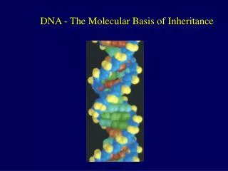
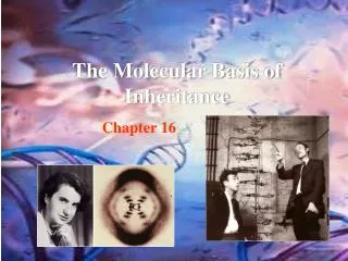
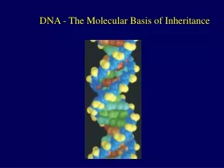
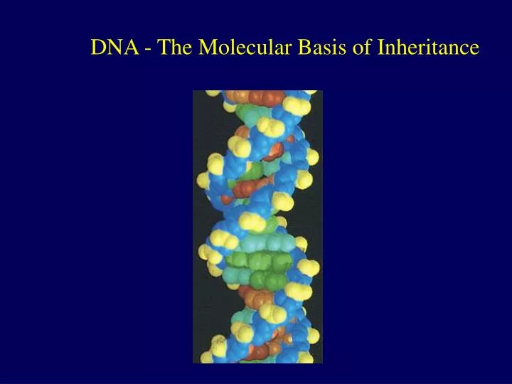
DNA - The Molecular Basis of Inheritance. James D. Watson & Francis H. Crick. In 1953 presented the double helix model of DNA Two primary sources of information: 1. Chargaff Rule: #A #T and #G #C. “A strange but possibly meaningless phenomenon”.

E N D
James D. Watson & Francis H. Crick • In 1953 presented the double helix model of DNA • Two primary sources of information: • 1. Chargaff Rule: #A#T and #G#C. “A strange but possibly meaningless phenomenon”. • 2. X-ray diffraction studies of Rosalind Franklin & Maurice H. F. Wilkins
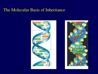
DNA Structure • Conclusion-DNA is a helical structure with distinctive regularities, 0.34 nm & 3.4 nm.
1962: Nobel Prize in Physiology and Medicine Watson, J.D. and F.H. Crick, “Molecular Structure of Nucleic Acids: A Structure for Deoxynucleic Acids”. Nature 171 (1953), p. 738. James D. Watson Francis H. Crick Maurice H. F. Wilkins What about? Rosalind Franklin
The Structure of DNA • DNA is composed of four nucleotides, each containing: adenine, cytosine, thymine, or guanine. • The amounts of A = T, G = C, and purines = pyrimidines [Chargaff’s Rule]. • DNA is a double-stranded helix with antiparallel strands [Watson and Crick]. • Nucleotides in each strand are linked by 5’-3’ phosphodiester bonds • Bases on opposite strands are linked by hydrogen bonding: A with T, and G with C.
5 end Hydrogen bond 3 end 1 nm 3.4 nm 3 end 0.34 nm 5 end The Basic Principle: Base Pairing to a Template Strand • The relationship between structure and function is manifest in the double helix • Since the two strands of DNA are complementary each strand acts as a template for building a new strand in replication • In DNA replication, the parent molecule unwinds, and two new daughter strands are built based on base-pairing rules
A A A A T A T T T T G C C C C G G G G C A T T T T A A A A T A A A A T A T T T T G G G G C G C C C C (a) The parent molecule has two complementary strands of DNA. Each base is paired by hydrogen bonding with its specific partner, A with T and G with C. (c) Each parental strand now serves as a template that determines the order of nucleotides along a new, complementary strand. (d) The nucleotides are connected to form the sugar-phosphate backbones of the new strands. Each “daughter” DNA molecule consists of one parental strand and one new strand. (b) The first step in replication is separation of the two DNA strands. DNA replication • The parent molecule unwinds, and two new daughter strands are built based on base-pairing rules T A G C A T T A C G
DNA Replication • DNA must replicate during each cell division • 3 alternative models for DNA replication were hypothesized: • Semiconservative replication • Conservative replication • Dispersive replication Conservative Dispersive Semi-conservative
Bacteria cultured in medium containing 15N Bacteria transferred to medium containing 14N Less dense DNA sample centrifuged after 20 min (after first replication) DNA sample centrifuged after 40 min (after second replication) More dense Second replication First replication Conservative model Semiconservative model Dispersive model Meselson-Stahl Experiments • Labeled the nucleotides of old strands with a heavy isotope of nitrogen (15N), new nucleotides were indicated by a lighter isotope (14N). • The first replication in the 14N medium produced a band of hybrid (15N-14N) DNA, eliminating the conservative model. • A second replication produced both light and hybrid DNA, eliminating the dispersive model and supporting the semiconservative model.
DNA Replication is “Semi-conservative” • Each 2-stranded daughter molecule is only half new • One original strand was used as a template to make the new strand
DNA Replication • The copying of DNA is remarkable in its speed and accuracy • Involves unwinding the double helix and synthesizing two new strands. • More than a dozen enzymes and other proteins participate in DNA replication • The replication of a DNA molecule begins at special sites called origins of replication, where the two strands are separated
Origin of replication Parental (template) strand 0.25 µm Daughter (new) strand 1 Replication begins at specific sites where the two parental strands separate and form replication bubbles. Bubble Replication fork 2 The bubbles expand laterally, as DNA replication proceeds in both directions. 3 Eventually, the replication bubbles fuse, and synthesis of the daughter strands is complete. Two daughter DNA molecules (a) In eukaryotes, DNA replication begins at many sites along the giant DNA molecule of each chromosome. In this micrograph, three replication bubbles are visible along the DNA of a cultured Chinese hamster cell (TEM). (b) Origins of Replication • A eukaryotic chromosome may have hundreds or even thousands of replication origins
DNA polymerase III adds nucleotides to primer DNA polymerase I degrades the RNA primer and replaces it with DNA Mechanism of DNA Replication • DNA replication is catalyzed by DNA polymerase III which needs an RNA primer • DNA polymerase III cannot initiate the synthesis of a polynucleotide, they can only add nucleotides to the 3 end • The initial nucleotide strand is an RNA primer • RNA primase synthesizes primer on DNA strand • DNA polymerase adds nucleotides to the 3’ end of the growing strand
New strand Template strand 5¢ end 3¢ end 5¢ end 3¢ end Sugar Base Phosphate DNA polymerase 3¢ end 3¢ end Pyrophosphate Nucleoside triphosphate 5¢ end 5¢ end Mechanism of DNA Replication • Nucleotides are added by complementary base pairing with the template strand • DNA always reads from 5’ end to 3’ end for transcription replication • During replication, new nucleotides are added to the free 3’ hydroxyl on the growing strand • The nucleotides (deoxyribonucleoside triphosphates) are hydrolyzed as added, releasing energy for DNA synthesis. • The rate of elongation is about 500 nucleotides per second in bacteria and 50 per second in human cells
3¢ 5¢ Parental DNA Leading strand 5¢ 3¢ Okazaki fragments Lagging strand 3¢ 5¢ DNA pol III Template strand Leading strand Lagging strand Template strand DNA ligase Overall direction of replication The Mechanism of DNA Replication • DNA synthesis on the leading strand is continuous • Only one primer is needed for synthesis of the leading strand • The lagging strand grows the same general direction as the leading strand (in the same direction as the Replication Fork). However, DNA is made in the 5’-to-3’ direction • Therefore, DNA synthesis on the lagging strand is discontinuous • For synthesis of the lagging strand, each fragment (Okazaki) must be primed separately, then DNA fragments are sythesized and subsequently ligated together
Overall direction of replication Lagging strand Leading strand Origin of replication Leading strand Leading strand Lagging strand OVERVIEW DNA pol III DNA ligase Replication fork 5¢ DNA pol I 3¢ Primase Parental DNA DNA pol III Primer 3¢ 5¢ Lagging strand Mechanism of DNA Replication • Many proteins assist in DNA replication • DNA helicases unwind the double helix, the template strands are stabilized by other proteins • Single-stranded DNA binding proteins make the template available • RNA primase catalyzes the synthesis of short RNA primers, to which nucleotides are added. • DNA polymerase III extends the strand in the 5’-to-3’ direction • DNA polymerase I degrades the RNA primer and replaces it with DNA • DNA ligase joins the DNA fragments into a continuous daughter strand
Enzymes in DNA replication Primase adds short primer to template strand Helicase unwinds parental double helix Binding proteins stabilize separate strands Ligase joins Okazaki fragments and seals other nicks in sugar-phosphate backbone DNA polymerase I (Exonuclease) removes RNA primer and inserts the correct bases DNA polymerase III binds nucleotides to form new strands
Replication 3’ 5’ 3’ 5’ 3’ 5’ 3’ 5’ Helicase protein binds to DNA sequences called origins and unwinds DNA strands. Binding proteins prevent single strands from rewinding. Primase protein makes a short segment of RNA complementary to the DNA, a primer.
Replication Overall direction of replication 3’ 3’ 5’ 5’ 3’ 5’ 3’ 5’ DNA polymerase III enzyme adds DNA nucleotides to the RNA primer.
Replication Overall direction of replication 3’ 5’ 3’ 5’ 3’ 3’ 5’ 5’ DNA polymerase proofreads bases added and replaces incorrect nucleotides.
Replication Overall direction of replication 3’ 3’ 5’ 5’ 3’ 5’ 3’ 5’ Leading strand synthesis continues in a 5’ to 3’ direction.
Replication Overall direction of replication 3’ 3’ 5’ 5’ Okazaki fragment 3’ 3’ 5’ 5’ 3’ 5’ Leading strand synthesis continues in a 5’ to 3’ direction. Discontinuous synthesis produces 5’ to 3’ DNA segments called Okazaki fragments.
Replication Overall direction of replication 3’ 3’ 5’ 5’ Okazaki fragment 3’ 3’ 5’ 5’ 3’ 5’ Leading strand synthesis continues in a 5’ to 3’ direction. Discontinuous synthesis produces 5’ to 3’ DNA segments called Okazaki fragments.
Replication Overall direction of replication 3’ 3’ 5’ 5’ Okazaki fragment 3’ 5’ 3’ 5’ 3’ 5’ Leading strand synthesis continues in a 5’ to 3’ direction. Discontinuous synthesis produces 5’ to 3’ DNA segments called Okazaki fragments.
Replication 3’ 5’ 3’ 5’ 3’ 5’ 3’ 3’ 5’ 3’ 5’ 5’ Leading strand synthesis continues in a 5’ to 3’ direction. Discontinuous synthesis produces 5’ to 3’ DNA segments called Okazaki fragments.
Replication 3’ 5’ 3’ 5’ 3’ 5’ 3’ 3’ 5’ 3’ 5’ 5’ Leading strand synthesis continues in a 5’ to 3’ direction. Discontinuous synthesis produces 5’ to 3’ DNA segments called Okazaki fragments.
5’ Replication 3’ 5’ 3’ 5’ 3’ 5’ 3’ 3’ 3’ 5’ 5’ Exonuclease activity of DNA polymerase I removes RNA primers.
Replication 3’ 3’ 5’ 3’ 5’ 3’ 3’ 5’ 5’ Polymerase activity of DNA polymerase I fills the gaps. Ligase forms bonds between sugar-phosphate backbone.
Overall direction of replication Lagging strand Leading strand Origin of replication Leading strand Leading strand Lagging strand OVERVIEW DNA pol III DNA ligase Replication fork 5¢ DNA pol I 3¢ Primase Parental DNA DNA pol III Primer 3¢ 5¢ Lagging strand Replication Fork Overview
Other Proteins That Assist DNA Replication • Helicase, topoisomerase, single-strand binding protein are all proteins that assist DNA replication
Proofreading • Mistakes during the initial pairing of template nucleotides and complementary nucleotides occur at a rate of one error per 100,000 base pairs. • DNA polymerase proofreads each new nucleotide against the template nucleotide as soon as it is added and can correct errors • If there is an incorrect pairing, the enzyme removes the wrong nucleotide and then resumes synthesis. • Mismatched nucleotides that are missed by DNA polymerase or mutations that occur after DNA synthesis is completed can often be repaired
Mutations • Mismatch repair: ‘wrong’ inserted base can be removed • Excision repair: DNA may be damaged by chemicals, radiation, etc. Mechanism to cut out and replace with correct bases • Each cell continually monitors and repairs its genetic material, with 100 repair enzymes known in E. coli and more than 130 repair enzymes identified in humans. • The final error rate is only one per ten billion nucleotides • Because the human genome is so large, even at this rate, mutations add up. Each of us probably inherited 3-4 mutations!
1 2 4 A thymine dimer distorts the DNA molecule. A nuclease enzyme cuts the damaged DNA strand at two points and the damaged section is removed. Nuclease DNA polymerase Repair synthesis by a DNA polymerase fills in the missing nucleotides. 3 DNA ligase DNA ligase seals the Free end of the new DNA To the old DNA, making the strand complete. Proofreading and Repairing DNA • DNA polymerases proofread newly made DNA, replacing any incorrect nucleotides • In mismatch repair of DNA, repair enzymes correct errors in base pairing • In nucleotide excision DNA repair nucleases cut out and replace damaged stretches of DNA
Accuracy of DNA Replication • The chromosome of E. coli bacteria contains about 5 million bases pairs • Capable of copying this DNA in less than an hour • The 46 chromosomes of a human cell contain about 6 BILLION base pairs of DNA!! • Printed one letter (A,C,T,G) at a time…would fill up over 900 volumes of Campbell. • Takes a cell a few hours to copy this DNA • With amazing accuracy – an average of 1 per billion nucleotides
5 Leading strand Lagging strand End of parental DNA strands 3 Last fragment Previous fragment RNA primer 5 Lagging strand 3 Primer removed but cannot be replaced with DNA because no 3 end available for DNA polymerase Removal of primers and replacement with DNA where a 3 end is available 5 3 Second round of replication 5 New leading strand 3 New lagging strand 5 3 Further rounds of replication Shorter and shorter daughter molecules Replicating the Ends of DNA Molecules • The ends of eukaryotic chromosomal DNA get shorter with each round of replication
1 µm Telomeres • Eukaryotic chromosomal DNA molecules have at their ends nucleotide sequences, called telomeres, that postpone the erosion of genes near the ends of DNA molecules
Telomerases • If the chromosomes of germ cells became shorter in every cell cycle essential genes would eventually be missing from the gametes they produce • An enzyme called telomerase catalyzes the lengthening of telomeres in germ cells