Download
1 / 32
330 likes | 522 Views
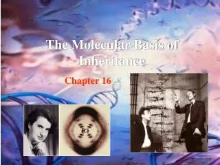
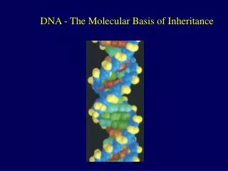
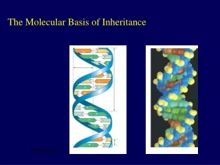
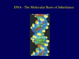
The Molecular Basis of Inheritance. Chapter 16. Life’s Operating Instructions. In 1953, James Watson and Francis Crick introduced a double-helical model for the structure of deoxyribonucleic acid, or DNA Hereditary information is encoded in DNA and reproduced in all cells of the body

E N D
The Molecular Basis of Inheritance Chapter 16
Life’s Operating Instructions • In 1953, James Watson and Francis Crick introduced a double-helical model for the structure of deoxyribonucleic acid, or DNA • Hereditary information is encoded in DNA and reproduced in all cells of the body • This DNA program directs the development of biochemical, anatomical, physiological, and (to some extent) behavioral traits
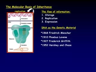
The Search for the Genetic Material: Scientific Inquiry • The role of DNA in heredity was first discovered by studying bacteria and the viruses that infect them • The discovery of the genetic role of DNA began with research by Frederick Griffith in 1928 • Griffith worked with two strains of a bacterium, one pathogenic and one harmless
The Search for the Genetic Material: Scientific Inquiry • When he mixed heat-killed remains of the pathogenic strain with living cells of the harmless strain, some living cells became pathogenic • He called this phenomenon transformation, now defined as a change in genotype and phenotype due to assimilation of foreign DNA
Can a genetic trait be transferred between different bacterial strains? Mixture ofheat-killedS cells andliving R cells Living S cells(control) Living R cells(control) Heat-killedS cells(control) Mouse dies Mouse healthy Mouse healthy Mouse dies Living S cells
Evidence That Viral DNA Can Program Cells • More evidence for DNA as the genetic material came from studies of viruses that infect bacteria • Such viruses, called bacteriophages(or phages), are widely used in molecular genetics research
Evidence That Viral DNA Can Program Cells Bacterialcell
Building a Structural Model of DNA: Scientific Inquiry • After DNA was accepted as the genetic material, the challenge was to determine how its structure accounts for its role in heredity • Maurice Wilkins and Rosalind Franklin were using a technique called X-ray crystallography to study molecular structure • Franklin produced a picture of the DNA molecule using this technique
Building a Structural Model of DNA: Scientific Inquiry Rosalind Franklin Franklin’s X-ray diffractionphotograph of DNA
Building a Structural Model of DNA: Scientific Inquiry • Franklin’s X-ray crystallographic images of DNA enabled Watson to deduce that DNA was helical • The X-ray images also enabled Watson to deduce the width of the helix and the spacing of the nitrogenous bases • The pattern in the photo suggested that the DNA molecule was made up of two strands, forming a double helix
Building a Structural Model of DNA: Scientific Inquiry • Watson and Crick built models of a double helix to conform to the X-rays and chemistry of DNA • Franklin had concluded that there were two outer sugar-phosphate backbones, with the nitrogenous bases paired in the molecule’s interior • Watson built a model in which the backbones were antiparallel (their subunits run in opposite directions)
Building a Structural Model of DNA: Scientific Inquiry • Watson and Crick reasoned that the pairing was specific, dictated by the base structures • They determined that adenine (A) paired only with thymine (T), and guanine (G) paired only with cytosine (C) • The Watson-Crick model explains: in any organism the amount of A = T, and the amount of G = C
Building a Structural Model of DNA: Scientific Inquiry Sugar Sugar Adenine (A Thymine (T) Sugar Sugar Guanine (G Cytosine (C)
The Basic Principle: Base Pairing to a Template Strand • Since the two strands of DNA are complementary, each strand acts as a template for building a new strand in replication • In DNA replication, the parent molecule unwinds, and two new daughter strands are built based on base-pairing rules
The Basic Principle: Base Pairing to a Template Strand Parent molecule Separation ofstrands “Daughter” DNA molecules,each consisting of oneparental strand and onenew strand
The Basic Principle: Base Pairing to a Template Strand • Watson and Crick’s semiconservative model of replication predicts that when a double helix replicates, each daughter molecule will have one old strand (derived or “conserved” from the parent molecule) and one newly made strand • Competing models were the conservative model (the two parent strands rejoin) and the dispersive model (each strand is a mix of old and new)
DNA Replication: A Closer Look • The copying of DNA is remarkable in its speed and accuracy • More than a dozen enzymes and other proteins participate in DNA replication
Getting Started • Replication begins at particular sites called origins of replication, where the two DNA strands are separated, opening up a replication “bubble” • A eukaryotic chromosome may have hundreds or even thousands of origins of replication • Replication proceeds in both directions from each origin, until the entire molecule is copied
Getting Started • At the end of each replication bubble is a replication fork, a Y-shaped region where new DNA strands are elongating • Helicasesare enzymes that untwist the double helix at the replication forks • Single-strand binding proteins bind to and stabilize single-stranded DNA • Topoisomerasecorrects “overwinding” ahead of replication forks by breaking, swiveling, and rejoining DNA strands
Getting Started 3 Topoisomerase 5 3 5 3 Helicase Single-strand bindingproteins 5
Getting Started • DNA polymerases cannot initiate synthesis of a polynucleotide; they can only add nucleotides to the 3end • The initial nucleotide strand is a short RNA primer • The primer is short (5–10 nucleotides long), and the 3 end serves as the starting point for the new DNA strand
Synthesizing a New DNA Strand • Enzymes called DNA polymerases catalyze the elongation of new DNA at a replication fork • Most DNA polymerases require a primer and a DNA template strand • The rate of elongation is about 500 nucleotides per second in bacteria and 50 per second in human cells
Antiparallel Elongation • The antiparallel structure of the double helix affects replication • DNA polymerases add nucleotides only to the free 3end of a growing strand; therefore, a new DNA strand can elongate only in the 5to3direction
Proofreading and Repairing DNA • DNA polymerases proofread newly made DNA, replacing any incorrect nucleotides • In mismatch repair of DNA, repair enzymes correct errors in base pairing • DNA can be damaged by exposure to harmful chemical or physical agents such as cigarette smoke and X-rays; it can also undergo spontaneous changes • In nucleotide excision repair, a nuclease cuts out and replaces damaged stretches of DNA
Evolutionary Significance of Altered DNA Nucleotides • Error rate after proofreading repair is low but not zero • Sequence changes may become permanent and can be passed on to the next generation • These changes (mutations) are the source of the genetic variation upon which natural selection operates
A chromosome consists of a DNA molecule packed together with proteins • The bacterial chromosome is a double-stranded, circular DNA molecule associated with a small amount of protein • Eukaryotic chromosomes have linear DNA molecules associated with a large amount of protein • In a bacterium, the DNA is “supercoiled” and found in a region of the cell called the nucleoid
A chromosome consists of a DNA molecule packed together with proteins • Chromatin,a complex of DNA and protein, is found in the nucleus of eukaryotic cells • Chromosomes fit into the nucleus through an elaborate, multilevel system of packing
A chromosome consists of a DNA molecule packed together with proteins • Chromatin undergoes changes in packing during the cell cycle • At interphase, some chromatin is organized into a 10-nm fiber, but much is compacted into a 30-nm fiber, through folding and looping • Though interphase chromosomes are not highly condensed, they still occupy specific restricted regions in the nucleus
A chromosome consists of a DNA molecule packed together with proteins • Most chromatin is loosely packed in the nucleus during interphase and condenses prior to mitosis • Loosely packed chromatin is called euchromatin • During interphase a few regions of chromatin (centromeres and telomeres) are highly condensed into heterochromatin • Dense packing of the heterochromatin makes it difficult for the cell to express genetic information coded in these regions