Download
1 / 27
290 likes | 620 Views
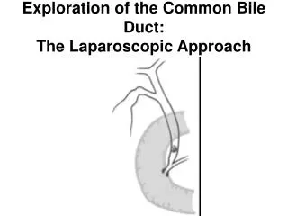
Revised AJCC Classification of Extrahepatic Bile Duct Tumors . Definitions. Anatomy of the Bile Ducts. left hepatic duct. right hepatic duct. hepatic hilum. perihilar bile ducts. proximal common duct (proximal hepatic duct). cystic duct. middle common duct. common bile duct.

E N D
Revised AJCC Classification of Extrahepatic Bile Duct Tumors
Definitions Anatomy of the Bile Ducts left hepatic duct right hepatic duct hepatic hilum perihilar bile ducts proximal common duct (proximal hepatic duct) cystic duct middle common duct common bile duct distal bile duct distal common duct ampulla of Vater 7th AJCC classification
Definitions AJCC Classification • Including bile duct tumors arising from the hepatic hilum of the right and left hepatic ducts to the distal common duct above the ampulla of Vater in the 6th edition (2002) • Separated into perihilar bile duct and distal bile duct tumors in the 7th edition (2010)
6th AJCC Classification Extrahepatic Bile Duct Tumor
7th AJCC Classification Definitions of TNM
7th AJCC Classification Definitions of TNM
7th AJCC Classification Anatomic Stage/Prognostic Groups
Summary Perihilar Bile Duct Tumors • T1: confined to the bile duct T2: beyond the wall of the bile duct wall • T2b: invasion of adjacent hepatic parenchyma (T3 in the 6th edition) • T3: unilateral vascular invasion • T4: bilateral biliary and/or vascular invasion • Regional lymph node metastasis: reclassified as stage IIIB (stage IIB in the 6th edition) • T4: unresectable based on local invasion (IVA) or distant disease (IVB)
Summary Distal Bile Duct Tumors • T1: confined to the bile duct T2: beyond the wall of the bile duct wall • T3: includes adjacent organs (invasion of adjacent organs such as the colon, stomach, duodenum or abdominal wall: classified as T4 in the 6th edition) • Invasion of the portal vein and hepatic artery: excluded in classification (T3 or T4 in the 6th edition) • No criteria on the longitudinal involvement
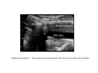
Radiologic Staging Perihilar Bile Duct Tumors • CT: wall thickening from left hepatic duct to distal common duct • Surgery: left major hepatobiliary resection, pathology: T1N0M0 • Major surgery was performed due to extensive longitudinal involvement
Radiologic Staging Perihilar Bile Duct Tumors • CT: wall thickening from both hepatic ducts to proximal common duct, periductal invasion (+) • Op: extended left hemihepatectomy, pathology: T2aN0M0
Radiologic Staging Perihilar Bile Duct Tumors • CT: wall thickening and mass invading to adjacent hepatic parenchyma, from left hepatic duct to hepatic hilum • Surgery: extended left hemihepatectomy, pathology: T2bN0M0 • T3 in the 6th edition, T2b in the 7th edition
Radiologic Staging Perihilar Bile Duct Tumors • CT: polypoid mass from both hepatic ducts to hepatic hilum, invasion to right hepatic artery • Surgery: segmental resection due to old age, pathology: T3N0M0 • Upstaged from stage IIA (T3N0M0) to stage IIIA (T3N0M0) in case of unilateral vascular invasion
Radiologic Staging Perihilar Bile Duct Tumors • CT: wall thickening from hepatic hilum to proximal common duct, lymph node (+) • Surgery: segment resection and lymph node dissection, pathology: T2aN1Mo • Upstaged from stage IIB (T2N1M0) to IIIB (T2aN1M0) due to regional lymph node metastasis
Radiologic Staging Distal Bile Duct Tumors • CT: polypoid mass from mid to distal, pericholedochal invasion (-) • Surgery: PPPD, pathology: T1N0M0 • CT: wall thickening from mid to distal, pericholedochal invasion (+) • Surgery: PPPD, pathology: T2N0M0
Radiologic Staging Distal Bile Duct Tumors • CT: wall thickening involving distal common duct, pancreatic invasion (+) • Surgery: PPPD and lymph node dissection, pathology: T3N0M0 • Pancreatic invasion: T3 in 6th and 7th editions
Radiologic Staging Distal Bile Duct Tumors • CT: wall thickening from distal common duct, pancreatic and duodenal invasion (+) • Surgery: PPPD and lymph node dissection, pathology: T3N0M0 • Downstaged from stage IV (T4N0M0) to stage IIA (T3N0M0) due to duodenal invasion
Radiologic Staging Distal Bile Duct Tumors • CT: polypoid mass from mid to distal common duct, pancreatic invasion (+), regional lymph node (+) • Surgery: PPPD, pathology: T3N1M0
Radiologic Staging Distal Bile Duct Tumors • CT: wall thickening from proximal to distal common duct, duodenal and pancreatic invasion (+), regional lymph node (+) • Surgery: PPPD, pathology: T3N1M0 • Downstaged from stage IV (T4N1M0) to stage IIB (T3N1M0) due to duodenal invasion
Radiologic Staging Distal Bile Duct Tumors • CT: wall thickening from middle to distal common duct, pancreatic invasion (+), regional lymph node (+) • Surgery: PPPD and portal vein resection, pathology: T3N1M0 • Portal vein invasion: T4 in the 6th edition, not defined T3 in the 7th edition (maybe T3 as other adjacent organ)
Limitations Perihilar Bile Duct Tumors • Invasion to hepatic parenchyma: CT (-), pathology (+)
Limitations Perihilar Bile Duct Tumors • Invasion to right hepatic artery: CT (+), pathology (-)
Limitations Perihilar Bile Duct Tumors • CT: Pancreatic invasion (+) • CT: Gallbladder invasion (+) • T stage of pancreatic and gallbladder invasion: not clearly defined
Limitations Distal Bile Duct Tumors • Pancreatic invasion: CT (-), pathology (+)
Limitations Distal Bile Duct Tumors • Invasion beyond bile duct wall: CT (+), pathology (-)
Limitations Distal Bile Duct Tumors • Mainportal vein invasion: CT (-), pathology (+) • Main portal invasion: T4 in the 6th edition, not clearly defined in the 7th edition (maybe T3 as other adjacent organ)
New Staging PerihilarCholangiocarcinoma DeOliveira MI, et al. Hepatology 2011;53:1363-1371