Download
1 / 133
1.38k likes | 1.75k Views
Che cosa è un momento magnetico di spin. SPIN S=1/2. S= [ S(S+1)] ½ h/2 p. Momento angolare di spin. m = - g S. Momento magnetico di spin. Quantizzazione.

E N D
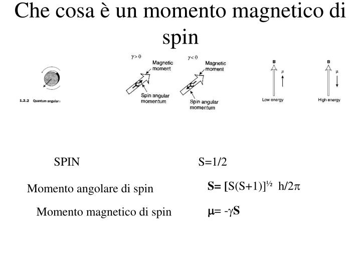
Che cosa è un momento magnetico di spin SPIN S=1/2 S= [S(S+1)]½ h/2p Momento angolare di spin m= -gS Momento magnetico di spin
Quantizzazione Il concetto di spin è associato alla meccanica quantistica, ovvero alla fisica che descrive il comportamento dei sistemi con particelle “infinitamente piccole” ovvero non trattabili con le regole della fisica classica. In accordo alla meccanica quantistica, l’energia della interazione tra il momento magnetico di spin ed il campo magnetico non puo’ essere “qualsiasi”, ma puo’ assumere solo valori definiti NO
Quantizzazione Il concetto di spin è associato alla meccanica quantistica, ovvero alla fisica che descrive il comportamento dei sistemi con particelle “infinitamente piccole” ovvero non trattabili con le regole della fisica classica. In accordo alla meccanica quantistica, l’energia della interazione tra il momento magnetico di spin ed il campo magnetico non puo’ essere “qualsiasi”, ma puo’ assumere solo valori definiti E= -mB cosq = -g[S(S+1)]1/2ħ B cosq E= = -għ m B m=+1/2 , -1/2
Quantizzazione Quindi, quando uno particella con uno spin S=1/2 è immersa in un campo magnetico, ci sono solo due valori di Energia possibili (permessi) E= +1/2ħg B E= -1/2ħg B E= -mB cosq = -g[S(S+1)]1/2ħ B cosq E= = -ħg m B m=+1/2 , -1/2
NMR-active nuclei with different spin numbers I=1/211H, 136C, 3115P, 199F, 157N I=3/22311Na3517Cl I=5/2178O 2713Al
Rapporto giromagnetico E= = -ħg m B DE= =ħg B La separazione in energia dipende dal valore del rapporto giromagnetico
B0 La frequenza di precessione di Larmor Ogni momento magnetico precede intorno al campo magnetico statico B0 Spin equivalenti precedono tutti con la stessa velocità w0 anche se ciascuno con una fase diversa a: m = +½ ma = +½g Ea = -½gB0 E = -mB0 E = -gmB0 equivalenti significa nuclidi di atomi identici b: m = -½ mb = -½g Eb = +½gB0 CH3CH2OH
I parametri NMR Il chemical shift Le costanti di accoppiamento La intensità dei segnali
Frequenza di precessione n0 = - g B0 /2π Se cosi fosse, ogni nucleo attivo entrerebbe in risonanza con il campo esterno alla sua frequenza e tutti gli isotopi uguali si comporterebbero allo stesso modo (un unico segnale). La frequenza di precessione di un determinato nucleo ad un determinato campo magnetico è detta FREQUENZA DI PRECESSIONE DI LARMOR Es: al campo magnetico di 11.7 T, La FREQUENZA DI PRECESSIONE DI LARMOR del nuclide 1H è 500 MHz.
Costante di schermo n= g/2p B0 (1-s) s = Costante di schermo Dipende dall’intorno elettronico
Chemical shift (n-nref/nref)*106 = d (ppm) Es: w1= 500.131 MHz w0=500.13 MHz w1-w0=1000 Hz = 1000/500.13x106 (ppm)= 2.0
TMS (Tetramethylsilane) chemical shift d d = 0
Fattori che influenzano il chemical shift Caratteristiche funzionaliEffetti attraverso lo spazioEffetti paramagnetici
Fattori che influenzano il chemical shift Caratteristiche funzionaliEffetti attraverso lo spazioEffetti paramagnetici
Simple shielding effects--electronegativity The amount of shielding the nucleus experiences will vary with the density of the surrounding electron cloud If a 1H nucleus is bound to a more electronegative atom e.g. N or O as opposed to C, the density of the electron cloud will be lower and it will be less shielded or “deshielded”. These considerations extend beyond what is directly bonded to the H atom as well. more electron withdrawing-- less shielded less electron withdrawing-- more shielded N C H H
Simple shielding effects--electronegativity less shielded higher resonance frequency more shielded lower resonance frequency aliphatic/alpha/beta etc.(HC) amides (HN) most HN nuclei come between 6-11 ppm while most HC nuclei come between -1 and 6 ppm.
More complex shielding effects:Aromatic protons aromatic region (6-8 ppm) amide region (7-10 ppm) One consequence of these effects is that aromatic protons, which are attached to aromatic rings, are deshielded relative to other HC protons. In fact, aromatic ring protons overlap with the amide (HN) region.
Amino acid structures and chemical shifts note: the shifts are somewhat different from the previous page because they are measured on the free amino acids, not on amino acids within peptides
“Average” or “random coil” chemical shifts in proteins One reason for this dispersion is that the side chains of the 20 amino acids are different, and these differences will have some effect on the Ha shift. The table at right shows “typical” values observed for different protons in the 20 amino acids. These were measured in unstructured peptides to mimic the environment experienced by the proton averaged over essentially all possible conformations. These are sometimes called “random coil” shift values. Note that the Ha shifts range from ~4-4.8, but Ha shifts in proteins range from ~3 to 5.5. So this cannot entirely explain the observed dispersion.
so you can tell if your protein is folded or not by looking at the 1D spectrum... poorly dispersed methyls poorly dispersed amides poorly dispersed alphas poorly dispersed aromatics unfolded ubiquitin very shielded methyl folded ubiquitin
A simple reason for the increased shift dispersion is that the environment experienced by 1H nuclei in a folded protein (B) is not the same as in a unfolded, extended protein or “random coil” (A). shift of particular proton in unfolded protein is averaged over many fluctuating structures will be near random coil value shift of particular proton in folded protein influenced by groups nearby in space, conformation of the backbone, etc. Not averaged among many structures because there is only one folded structure. So, some protons in folded proteins will experience very particular environments and will stray far from the average.
13C NMR The rules discussed for 1H spins, (shielding and deshielding effects) hold also for 13C spins. Some general features of 13C should be pointed out: Unlike 1H atoms, 13C atoms may form a different number and type of chemical bonds. Therefore, the shielding/deshielding effects are much more effective. The chemical shift range of 13C spins spans more than 200 ppm
A protein 13C NMR spectrum (low resolution) Aromatic signals Aliphatic Backbone CO and side chain COO- signals
13C NMR The rules discussed for 1H spins, (shielding and deshielding effects) hold also for 13C spins. Some general features of 13C should be pointed out: The amino acid dependence of chemical shift values is stronger for 13C atoms than in 1H atoms. Therefore, each amino acid has an almost unique pattern of 13C chemical shifts
I parametri NMR Il chemical shift Le costanti di accoppiamento La intensità dei segnali Il rilassamento
Accoppiamento scalare 2J 3J 3J
Accoppiamento scalare 1JHC 1JCC 1JHN
Accoppiamento scalare 13C 1H
Accoppiamento scalare 13C 1H
Accoppiamento scalare b b a a 13C 1H I S
Accoppiamento scalare omonucleare 3J HNHa 2J HbHb
Accoppiamento scalare 2J 3J 3J