Download
1 / 21
210 likes | 474 Views
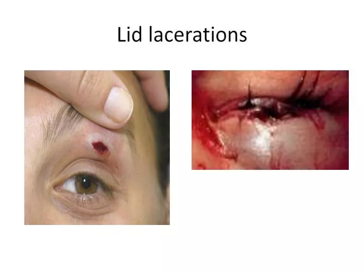
Lid lacerations. Ecchymosis. m anagement of uncomplicated lid abrasions. managed in office with: cleansing antibiotic ung dressing wound. management of deeper or canalicular lacerations. refer for surgical repair. management of ecchymosis. cold compresses for first 24-48 hours

E N D
management of uncomplicated lid abrasions • managed in office with: • cleansing • antibiotic ung • dressing wound
management of deeper or canalicular lacerations • refer for surgical repair
management of ecchymosis • cold compresses for first 24-48 hours • followed by warm compresses
Insect Bites—treat with antihistamine and/or oral steroid (medrol dose pack)
management of corneal abrasions that is limited to epithelia • observe for fluorescein staining • abrasion vs ulcer • antibiotic ung or gtts • NSAID and/or cycloplegic for pain • bandage CL or patching • rest • follow-up next day if patched; 3 days if superficial
corneal foreign body management • remove foreign body (unless it penetrates into globe) • antibiotic ung, cycloplegic, NSAID • follow-up 1 day; monitor for secondary infection or inflammation in organic or stone foreign bodies. • metallic foreign bodies need to be removed to prevent secondary infection or ulceration
Seidel’s sign Name the sign?
Iris prolapse (pupil points where iris bulges out of cornea)
management of penetrating corneal injuries • determine if it is an open or closed globe injury • Seidel test • check for cornea edema • check for anterior chamber depth • closed globe • topical antibiotic • cycloplegia • bandage CL • open globe • shelved type: treat as closed type • larger penetrating type: eye shield & refer
management of ocular chemical injury • DO NOT WASTE TIME ASSESSING THE EYES! • immediately flush, flush, flush the eye (30 minutes plus!) • alkaline burnes are MUCH WORSE than acid burns • antibiotics, lubrication, cycloplegia, topical steroids
management of hyphema • good history • blood dyscrasias • bleeding disorders • current anticoagulant therapy • grade the hyphema • grade 1: < 1/3 of AC • grade 2: 1/3 to 1/2 of AC • grade 3: ½ to less than full • grade 4: total (“8 ball”) • check IOP; manage if elevated • treat with: • cycloplegia • topical steroids • eye shield • limit activity; elevate head while reclined
management of orbital hemorrhage • observe signs of proptosis, EOM restriction, resistance to retropulsion, increased IOP and central retinal artery pulsation • vision threatening: • treat CRAO & refer to surgeon for orbital decompression • non-vision threatening: • CT of orbits • cold compresses, head elevation, manage IOP as needed
List orbital fractures in order of most common to least common: • floor & medial wall • roof • lateral wall
Ocular signs of orbital fracture • motility restriction • enophthalmos • crepitus • hypoesthesia of the orbital rim involved • (Note cornea light reflexes in photo—left eye has orbital fracture)
management of orbital fractures • scan or don’t scan • nasal decongestants • ice packs • oral analgesics & oral antibiotics • refrain from nose blowing, coughing & sneezing • surgery usually done after swelling goes down (~2 weeks)