Download
1 / 41
410 likes | 561 Views
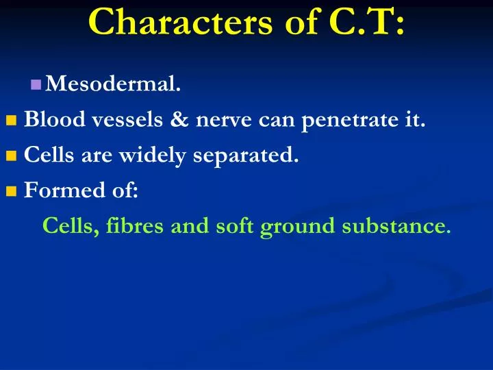
Characters of C.T:. Mesodermal. Blood vessels & nerve can penetrate it. Cells are widely separated. Formed of: Cells, fibres and soft ground substance. Fixed - Fibroblast. -Fixed macrophages. - Fat adipose cells. -U.M.C. - Pericytes. -Endothelial cells. Reticular cells.

E N D
Characters of C.T: • Mesodermal. • Blood vessels & nerve can penetrate it. • Cells are widely separated. • Formed of: Cells, fibres and soft ground substance.
Fixed • - Fibroblast. • -Fixed macrophages. • - Fat adipose cells. • -U.M.C. • - Pericytes. • -Endothelial cells. • Reticular cells. • *stable • *long lived • *non motile • New classification Free 1- Free macrophages. 2-Plasma cells. 3-Mast cell. 4-Plasma cell. 5-Leukocytes. * Non stable *short lived *motile
Classification of C.T. Fibres: 1-White collagenus bundle. 2-Yellow elastic fibres. 3-Reticular fibres.
(c) Different types of C.T. proper: 1-Loose areolar C.T. 2-White fibrous C.T. 3-Elastic C.T. 4-Reticular C.T. 5-Mucoid C.T. 6-Fatty adipose C.T.
3- Histiocytes, Macrophages or Phagocytes: • They are irregular cells with blunt long processes, Macrophages can be demonstrated by vital staining
Function: • They engulf dead neutrophilic leucocytes and bacteria by a phagocytes surrounding the object forming multinucleated foreign body giant cell. • Marcophages increase in number in chronic inflammation by: 1- Multiplication of already present macrophages.
4-Pigment Cells or Melanocytes • They are small branching cells rich in melanin pigments with a small centrally situated nucleus. • They are found in the skin, eye (retina, iris and choroid) and in substantianigra of midbrain.
Function: • They carry melanin pigment which protect the body from the injurious effect of the sun.
B- Rounded C.T. Cells 1-Plasma cells • They are irregularly ovoid in shape, small and centrically nucleus. • The nuclear chromatin is concentrated towards the nuclear membrane in a regular manner giving it a “cartwheel” appearance.
The cytoplasm is basophilic with a pale area representing Golgi apparatus “Negative Golgi image”. • The cytoplasm of the plasma cells contains acidophilic inclusions known as RUSSEL BODIES.
Functions: • Plasma cells are originated from B-lymphocytes: • B-lymphocytes antigen_plasmablasts plasma cells. • Plasma cells produce the circulating antibodies and they are responsible for immune response.
2-Mast Cells • They are large ovoid cells with ovoid nuclei. • Cytoplasm is full of basophilic granules. • These granules are water soluble and are metachromatically stained by toluidine blue, so they are stained purple • Mast cells are present in loose C.T. in relation to blood vessels.
Functions: • -Secrete heparin (anticoagulant), • -Secrete histamine, which participates in antigen antibody reaction. • -May have a role in serotonin secretion which, is a vasoconstrictor substance.
3- Fat Cells • They are rounded or oval cells with flattened peripheral nuclei and thin rim of cytoplasm, containing large fat droplets. • In H&E sections, the fat dissolved leaving an empty space and giving the appearance of a “signet”. • The fat cells can be stained orange with Sudan III and black with Sudan black.
Function: • They support vital organs as the kidney. • They store fat, which is the main source of energy. • They decrease loss of heat from the skin.
Classification of C.T. fibres: 1-White collagen us bundle. 2-Yellow elastic fibres. 3-Reticular fibres.
A-White or Collagen Fibers • About1-20 µm formed of thick backed fibres forming groups of bundles closely arranged together. • Collagenous bundles are elastic -long- cylindrical appearance. • Mainly locatedin • Tendon. • Joint capsule. • Deep fascia.
B-Yellow or Elastic Fibres -They are thinner than collagen fibres, refractile.can branch, anastmose and are highly elastic. -They occur as fenestrated lamellae in arteries, and they can be stained brown with orcein, blue with Mallory and yellow with van Geison.
C-Reticular Fibres -They are fine branching fibres stain brownish black with silver. • Function: • They participate in the stroma formation of • many organs as the kidney, liver and lung.
1-Loose Areolar C.T. -It contains all the elements of C.T. -Cells mainly fibroblasts and histiocytes. Fibres mainly collagen and ground substance. found in : -Dermis of skin. -Adventitia of blood vessels. -Under epithelial membranes.
Function: 1-It is important in exchanging nutrients to and from blood vessels. 2-It binds structures together. 3-It limits the spread of localized infection.
Mucoid C.T(Wharton’s Jelly): Presentmainly in the embryo. Formed of: Mucoid cells = U.M.C. or fibroblast joined by their processor. Matrix: jelly like rich in mucin thin collagenfibres. Sites: In embryo: umbilical cord. In adult: Vitreous humour of eye. Pulp of growing teeth.
Dense collagen C.T.: - Either regular or irregular. I-The regular dense collagenC.T is characterized by close packing of fibres is found in tendons,aponeuroses, ligaments and cornea of the eye. II-The irregular dense collagen C.T is found in dermis, capsules of some organs, perichondrium and sclera of the eye.
Yellow elastic C.T: Formed of condensed elastic fibres separated by fibroblast. Function: stretchable. Sites:Aorta & large arteries. Ranched tree. Vocal cords. Ligamentumnuchae (neck). Ligamentumflavum (between vertebrae).
Reticular C.T. -It is formed of reticular cells and fibres. The cells are stellate in shape with processes. -This tissue stains brownish black with silver -The main sites -Lymphoid tissues -Bone marrow -Stroma of body organs
Adipose C.T. -It is formed of lobes and lobules of fat cells separated by loose C.T. -It is richly supplied with blood vessels. -According to the vascularity and function, it is divided into white and brown adipose C.T. The white type (Unilocular)is distributed in : * Adults subcutaneous tissue and yellow bone marrow. -The brown type (Multilocular) is present : * Foetus and newborn infants.