Download
1 / 38

E N D
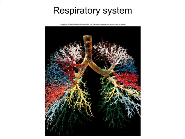
1. Respiratory system
2. What is respiration? Multiple definitions
Ventilation of lungs (breathing)
Exchange of gases between air and blood
Also between blood and tissues
Oxygen consumption during metabolic activities
Happens in what organelle?
3. What organs are involved? Conducting regions
Nose
Larynx
Epiglottis
Trachea
Bronchi
Lungs
Bronchioles
Respiratory region
Alveoli
Upper respiratory
Head and neck
Lower respiratory
Trachea to lungs
4. What does the nose do? Receive and humidify incoming air
Smells
Voice resonating chamber
Filter out bacteria
5. Why do I only breath out of one chamber at a time? Nasal fossa = chamber
Divided by nasal septum
Lamina propria of inferior concha swell
Every 30-60 minutes
Alternate chambers
Allows for rehydration
Epistaxis
Nose bleed; most common in inferior concha
6. What�s up with the pharynx and larynx? Pharynx extends from nose/mouth to larynx
Good at trapping large dust particles
Larynx = voice box
Job: keep food/water out of trachea
Also produces sound
Glottis (opening) guarded by epiglottis
7. What does the trachea do? Hyaline cartilage rings stiffen tube
Directs air to lungs via bronchi
Pseudostratified epithelium
Mucociliary escalator: cilia sweep mucus upward to pharynx
8. What parts make up the lungs? Right lung: superior, middle, inferior lobes
Left lung: superior and inferior lobes only
Bronchial tree
Branched air tubes from primary bronchi (two) to secondary bronchi to tertiary bronchi to bronchioles
Overlapping plates of cartilage give shape
9. What parts make up the lungs? Bronchioles (no cartilage) have smooth muscle to dilate/contract
Asthma
Bronchiolconstriction (irritants, cold air, histamine) and bronchioldilation
Click below for asthma movie!
10. What parts make up the lungs? Terminal bronchioles follow after bronchioles
Final branches of conducting division (region)
Cilia prevent congestion via mucociliary escalator
Next are respiratory bronchioles
Beginning of respiratory division
Divide into alveolar ducts
End in alveolar sacs
11. What do the alveoli do? Approx. 150 million = great S.A.
Mostly squamous alveolar cells�why?
A few cuboidal cells (great alveolar cells)
Secrete pulmonary surfactant
Disrupts hydrogen bonds
Prevents alveolar collapse during expiration
Premature infants lack
12. What do the alveoli do? Alveolar macrophages consume anything mucus doesn�t strain out
Items smaller than 2 micrometers
Capillaries surround each alveolus
Distance air must travel
Called respiratory membrane
Squamous alveolar cell, basement membrane, capillary squamous cell
13. What are the pleurae and why are they there? Moist serous membrane covering outside of lungs
Visceral pleura inside
Folds out at hillum to form outer parietal pleura
Pleural cavity: between parietal and visceral layers
Filled with pleural (serous) fluid
Pleurae functions
Reduce friction
Create pressure gradient to help with lung inflation
Compartmentalization: prevent infection spread to neighboring organs
14. How does breathing occur? Atmospheric pressure drives respiration
Atm. pressure = weight of air column
1 atm. = 760 mmHg
Boyle�s law: pressure and volume are inversely proportional
Intrapulmonary pressure changes opposite to volume load
Lower intrapulmonary pressure below 760 mmHg to draw breath
15. What happens during inspiration? Diaphragm does most of the work
Phrenic nerves cause diaphragm to flatten
External intercostals also contract, elevating (expanding) ribcage
Increases lung volume so pressure ______
Parietal pleurae cling to external intercostals
Visceral pleurae cling to parietal pleurae
Helps increase volume
16. What happens during inspiration? Air is warmed to assist volume expansion
500 ml air inhaled during resting inspiration
150 ml is dead air (held in conducting regions)
Thus only 350 ml to alveoli (respiratory division)
Alveolar ventilation rate (AVR) = 350 ml (or whatever value) X number of breath per minute
17. What happens during expiration? Passive process
Diaphragm and external intercostals allowed to relax
Natural elasticity of ribcage, lungs and tendons assist via recoil
Boyle�s law: volume decreases so pressure ___________ and air ______
Hard exhale: use internal intercostals to depress ribcage
18. Why does a lung collapse? Pneumothorax: air in the pleural cavity
Can happen if thoracic wall is punctured
Separates visceral and parietal pleurae
Result? Lung collapse = atelectasis
Can also happen if area of lung is blocked
Blood absorbs air and lung collapses
X-Ray shows complete atelectasis of the right lung. X-Ray shows complete atelectasis of the right lung.
19. How is ventilation measured? Spirometer
Measures respiratory volumes
Tidal volume: amount of air inhaled or exhaled when relaxed
Inspiratory reserve volume: amount can breath in above tidal volume with maximum effort
Expiratory reserve volume
Residual volume: what remains in lungs after maximum expiration (keeps alveoli inflated)
20. What are some breathing variations? Apnea: temporary breath cessation
Dyspnea: gasping, labored breathing, shortness of breath
Orthopnea: dyspnea while lying down
Eupnea: normal, quiet breathing
Hyperpnea: increased rate and depth of breathing
Tachypnea: accelerated respiration
Hyperventilation: expels too much CO2, raising pH
Hypoventilation: increases CO2, lower pH
21. What controls breathing? Medulla oblongata
Inspiratory neurons
Expiratory neurons (don�t fire during eupnea)
Unknown how rate of breathing is set
Pons
Regulates ventilation via pneumotaxic center
Sends inhibitory signals to med. Obl. inspiratory center
More impulses = shorter breath
Voluntary control: motor cortex of frontal lobe
Automatic controls can override to protect organism
Ondine's curse: person must remember to take each breath. Can�t sleep without the help of a mechanical ventilator. From German legend: Ondine (a water nymph) took a mortal lover. The lover was unfaithful to her and so king of nymphs placed curse: lover had to remember to take each breath. Couldn�t go to sleep or would suffocate. Died from suffocation due to exhaustion.Ondine's curse: person must remember to take each breath. Can�t sleep without the help of a mechanical ventilator. From German legend: Ondine (a water nymph) took a mortal lover. The lover was unfaithful to her and so king of nymphs placed curse: lover had to remember to take each breath. Couldn�t go to sleep or would suffocate. Died from suffocation due to exhaustion.
22. What happens during gas exchange? Air composition: O2, N2, H2O, CO2
Dalton�s law: partial pressure of each adds up to total pressure
What should they add up to at sea level?
23. What affects gas exchange? Takes about 0.25 secs to create equilibrium
Erythrocyte takes 0.75 sec to pass through alveolar cap.
During exercise erythrocyte present for 0.3 sec
24. What affects gas exchange? Concentration gradients of gases
Blood entering lungs
PO2 = 40 mmHg
PCO2 = 46 mmHg
Blood leaving lungs
PO2 = 95 mmHg
PCO2 = 40 mmHg
Solubility: Same amount of gases exchange, though, bec. CO2 is about 20 times as soluble in water
25. What affects gas exchange? Membrane thickness: respiratory membrane v. thin
Left ventricular failure = edema and thickened resp. memb.
Result: gases can�t equilibrate fast enough
Membrane area: more resp. memb. S.A. = better gas exchange
Emphysema, tuberculosis decrease S.A.
26. How is oxygen transported? Oxygen: 98% bound to hemoglobin
Review hemoglobin structure
How many molecules of oxygen can one hemoglobin hold?
As more oxygen bind to hemoglobin, affinity for oxygen increases
Oxyhemoglobin vs. deoxyhemoglobin
CO poisoning: carboxyhemoglobin (HbCO): competes with oxygen for binding site
Binds 210 times more tightly than oxygen to heme group
27. How is carbon dioxide transported? Three ways
Carbonic acid
70% of CO2 is hydrated
CO2 + H2O ? H2CO3 ? HCO3- + H+
Carbamino compounds (23%)
To plasma proteins and hemoglobin to form carboaminohemoglobin�HbCO2 (different from carboxyhemoglobin)
Binds to polypeptides, not to heme groups
Dissolved gas (7%)
Dissolves in plasma like CO2 in soda pop
28. What is carbon dioxide loading? Carbonic acid reaction occurs in plasma and erythrocytes
Carbonic anhydrase in RBCs speeds reaction
Chloride shift: HCO3 diffuse out of RBCs, replaced with Cl-
H+ binds to Hb (this buffers RBC pH)
29. What is oxygen unloading? H+ bound to HBO2 decreases affinity for O2
Causes RBC to offload oxygen more easily
Under what conditions would this happen?
Venous reserve: oxygen not offloaded
Can sustain life for up to five minutes
30. What happens in exhalation? Carbon dioxide offloading
Exact reverse of loading process
Oxygen loading
Reverse of offloading
31. Does Hb always offload the same amount of O2? No!
If PO2 of tissue is lower, more is offloaded
Higher temp = more offloaded
Bohr effect: active tissues put off more CO2 which lowers blood pH
This causes more O2 offloading = Bohr effect
BPG (bisphosphoglycerate) binds to Hb and promotes O2 offloading
RBS anaerobic fermentation intermediate
Fever, growth hormone, NE stimulate BPG synthesis
32. Does Hb always load the same amount of CO2? No!
Haldane effect: low HbO2 allows more CO2 transport
HbO2 doesn�t bind CO2 as well as deoxyhemoglobin (HHb)
HHb binds more H+ which removes H+ from solution
How does this affect the reaction CO2 + H2O ? H2CO3 ? HCO3- + H+?
33. How does blood chemistry affect ventilation? Peripheral chemoreceptors monitor
Carotid bodies and aortic bodies
Central chemoreceptors also monitor
In brainstem
Both monitor blood pH
How does this relate to O2 and CO2 levels?
34. How does blood chemistry affect ventilation? H+ in CSF is main site of monitoring
H+ doesn�t cross blood-brain barrier but CO2 does, then reacts with water
Problem: few proteins present to buffer H+
Acidosis: blood pH lower than 7.35
Common cause: hypercapnia (high CO2 value)
To correct: blow off CO2 rapidly (hyperventilation)
Alkalosis: above 7.45
Common cause: hypocapnia
To correct: take up CO2 rapidly (hypoventilation)
35. How does blood chemistry affect ventilation? Ketoacidosis: from diabetes mellitus
Lipid oxidation produces ketone bodies
Leads to low pH
Hyperventilation called Kussmaul respiration
Blowing off CO2 helps remove H+ and compensate for ketone body H+ production
36. What are some respiratory disorders? Hypoxia: oxygen deficiency
Hypoxemic hypoxia: low arterial PO2
High altitude, impaired pulmonary function, respiratory arrest, lung disease
Ischemic hypoxia: inadequate blood circulation
e.g. from congestive heart failure
Anemic hypoxia: from anemia
Histotoxic hypoxia: from metabolic poison (e.g. cyanide)
Hyperoxia: produces hydrogen peroxide and free radicals
37. What are some respiratory disorders? Chronic obstructive pulmonary disease (COPD)
Long-term obstruction of airflow
Asthma
Chronic bronchitis: cilia immobilized and reduced in number
More goblet cells present: produces sputum
Emphysema: alveolar walls destroyed
Smokers often have one or more of these
38. How does smoking and cancer affect the lungs? Lung cancer accounts for the most cancer-related deaths
Causes: smoking, followed by pollution
Tobacco: more than 15 carcinogens
Adam picture is a lateral view of a CXR in a patient with central cancer of the lung. Adam picture is a lateral view of a CXR in a patient with central cancer of the lung.


