Download
1 / 136
1.43k likes | 1.79k Views
RESPIRATORY SYSTEM. Organs of the Respiratory System. Nose Pharynx Larynx Trachea Bronchi Lungs—alveoli. Respiratory System Anatomy. Structurally divided into Upper respiratory system Above the larynx Nose, pharynx and associated structures Lower respiratory system Below the larynx

E N D
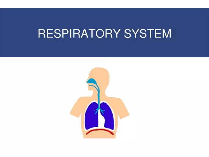
Organs of the Respiratory System • Nose • Pharynx • Larynx • Trachea • Bronchi • Lungs—alveoli
Respiratory System Anatomy • Structurally divided into • Upper respiratory system • Above the larynx • Nose, pharynx and associated structures • Lower respiratory system • Below the larynx • Larynx, trachea, bronchi and lungs
Respiratory tract • Functionally divided into • Conducting zone • conducts air to lungs • Nose, pharynx, larynx, trachea, bronchi, bronchioles and terminal bronchioles • Respiratory zone • main site of gas exchange • Respiratory bronchioles, alveolar ducts, alveolar sacs, and alveoli
Components of the Respiratory System Five Functions of the Respiratory System • Protects respiratory surfaces from outside environment • Participates in olfactory sense • Produces sounds • Moves air to and from exchange surfaces of lungs 5. Provides extensive gas exchange surface area between air and circulating blood
The Nose • Air enters the respiratory system through nostrils or external nares • Nasal cavity opens into nasopharynx through internal nares • Nasal hairs • Are in nasal vestibule • Are the first particle filtration system
Components of the Respiratory System The Respiratory Defense System • Filtration in nasal cavity removes large particles • Mucous cells and mucous glands produce mucus that bathes exposed surfaces • Cilia • Sweep debris trapped in mucus toward the pharynx (mucus escalator) • Alveolar macrophages engulf small particles that reach lungs
The Nose • The Nasal Cavity • The nasal septum divides nasal cavity into left and right • Mucous secretions from paranasal sinus and tears clean and moisten the nasal cavity • Superior portion of nasal cavity is the olfactory region provides sense of smell
The Nose • The Nasal Mucosa • Warms and humidifies inhaled air for arrival at lower respiratory organs • Breathing through mouth bypasses this important step
Paranasal Sinuses • Hollow portions (cavities) of bones surrounding the nasal cavity • Sinuses are located in the following bones • Frontal bone • Sphenoid bone • Ethmoid bone • Maxillary bone
Upper Respiratory Tract—Paranasal Sinuses Figure 13.2
Paranasal Sinuses • Function of the sinuses • Lighten the skull • Act as resonance chambers for speech • Produce mucus that drains into the nasal cavity • What is Sinusitis? • Sinusitis is the result of infection of these paranasal sinuses.
The Pharynx • Starts at internal nares and extends to cricoid cartilage of larynx • A chamber shared by digestive and respiratory systems • Contraction of skeletal muscles assists in deglutition
The Pharynx Divided into the • Nasopharynx • Oropharynx, • Laryngopharynx Functions of Pharynx • Passageway for air & food • Resonating chamber • Houses tonsils
The Larynx • A cartilaginous structure that surrounds the glottis, (narrow space between 2 vocal chords) • Air Flow -From the pharynx enters the larynx • the Larynx made of 3 large, unpaired cartilages
The Larynx Anterior Posterior • 1.Thyroid cartilage • 2. Cricoid cartilage • 3. Epiglottis
How do we produce speech? • Sound is produced by an air stream from the lungs, which goes through the trachea and the oral and nasal cavities. • Speech is produced by • Phonation • Articulation
How do we produce speech? • The phonation process occurs at the larynx. The larynx has two horizontal folds of tissue in the passage of air; they are the vocal chords. • The gap between these folds is called the glottis. • Sound is varied by Tension on vocal cords and Voluntary muscles
How do we produce speech? • Glottis - closed -no air can pass • Glottis - wide open, as in normal breathing, - the vibration of the vocal folds is reduced, producing the “voiceless sounds”. • Glottis - a narrow opening - make the vocal folds vibrate producing the “voiced sounds”.
How do we produce speech? • The articulation process takes place in the mouth and it is the process through which we can differentiate most speech sounds. • The oral cavity (upper and lower lips, upper and lower teeth, tongue and roof of the mouth) acts as a resonator, and the articulators • Paranasal sinuses- resonator
The Trachea • Also called the windpipe • Extends from the cricoid cartilage into mediastinum • Where it branches into right and left pulmonary bronchi
The Bronchial Tree • The Bronchial Tree – the primary bronchi and their branches • Primary bronchi • Secondary bronchi • Tertiary bronchi • Bronchioles • Terminal bronchioles • All but terminal bronchioles have reinforcing cartilage in their walls
The Bronchi • The Primary Bronchi • Right and left primary bronchi • The Right Primary Bronchus • Wider ( bigger diameter)/Shorter/ Descends at a steeper angle
The Lungs • The Lungs • Left and right lungs • Are in left and right pleural cavities • The base • Inferior portion of each lung rests on superior surface of diaphragm
The Lungs • Hilum ( The Root of the Lung) • Where pulmonary nerves, blood vessels, lymphatics enter lung • Anchored in meshwork of connective tissue in the mediastinum
The Lungs • The right lung has three lobes • Superior, middle, and inferior ; separated by oblique and horizontal fissures • The left lung has two lobes • Superior and inferior ; separated by an oblique fissure
Respiratory Zone Respiratory bronchioles Alveolar ducts Alveolar sacs Alveoli (air sacs) • Site of gas exchange = alveoli only
Alveoli • Alveoli- Cup-shaped outpouching • Alveolar sac – 2 or more alveoli sharing a common opening
Alveoli Respiratory Tissue.
Alveoli 2 types of alveolar epithelial cells • Type I alveolar cells – form nearly continuous lining, more numerous than type II, main site of gas exchange • Type II alveolar cells (septal cells) – free surfaces contain microvilli, secrete alveolar fluid (surfactant reduces tendency to collapse)
Alveoli • Surfactant • Is an oily secretion • Contains phospholipids and proteins • Coats alveolar surfaces and reduces surface tension
Respiratory Membrane Has three Layers • Squamous epithelial lining of alveolus (type I and type II alveolar cells) • Endothelial cells lining an adjacent capillary • Fused basement membrane (Basal laminae) between alveolar and endothelial cells Very thin – only 0.5 µm thick to allow rapid diffusion of gases
The Lungs • The Pleura • Consists of two layers • Parietal pleura • Visceral pleura • Pleural fluid • Lubricates space between two layers
Four Events of Respiration Respiratory gas transport Internal respiration External respiration
Four Events of Respiration • Pulmonary ventilation—moving air in and out of the lungs (= Breathing) • External respiration—gas exchange between pulmonary blood and alveoli • Oxygen is loaded into the blood • Carbon dioxide is unloaded from the blood 3. Respiratory gas transport—transport of oxygen and carbon dioxide via the bloodstream 4. Internal respiration—gas exchange between blood and tissue cells in systemic capillaries
Pulmonary Ventilation • Completely mechanical process that depends on volume changes in the thoracic cavity • Volume changes lead to pressure changes, which lead to the flow of gases to equalize pressure
Mechanics of Breathing Pulmonary ventilation (= Breathing) • Two phases • Inspiration ( inhalation) • flow of air into lungs • Always active • Expiration (exhalation) • air leaving lungs • Active or passive
Inhalation/ inspiration • Achieved by increasing size of lungs • lungs expand, increasing lung volume, decreasing pressure below atmospheric pressure • Boyle’s Law – pressure of a gas in a closed container is inversely proportional to the volume of the container • Pressure inside alveoli become lower than atmospheric pressure - External air is pulled into the lungs
Inspiration During inspiration external air is pulled into the lungs due to • Increase in intrapulmonary volume • Decrease in gas pressure
Inspiration • Inspiration is active process • Lungs expansion due to the Respiratory Muscles for Inspiration • The diaphragm • External intercostal muscles of the ribs • Accessory respiratory muscles: • activated when respiration increases significantly • Sternocleidomastoid • Serratus anterior • Pectoralis minor • Scalene muscles
Inspiration 1. Diaphragm • most important muscle of inhalation • Flattens, lowering dome when contracted • Contraction draws air into lungs • Responsible for 75% of air entering lungs during normal quiet breathing 2. External intercostals • Contraction elevates ribs • 25% of air entering lungs during normal quiet breathing 3. Accessory muscles for deep, forceful inhalation • Contraction elevates ribs