Download
1 / 80
800 likes | 970 Views
Honors Biology Ch. 12. Molecular Genetics. CH. 12 Molecular Genetics. I. DNA: The Genetic Material - DNA: The Chemical Basis of Heredity that forms the universal genetic code of cells. A. Discovery of the Genetic Material.

E N D
HonorsBiologyCh. 12 MolecularGenetics
CH. 12 Molecular Genetics • I. DNA: The Genetic Material • - DNA: The Chemical • Basis of Heredity that • forms the universal • genetic code of cells
A. Discovery of the Genetic Material • - In the early 1900’s scientists were debating what was the genetic material: • DNA vs. Protein.
1. Griffith • - In 1928 Griffith showed that a heat killed, but lethal strain of pneumonia bacteria could ‘transform’ a harmless strain of pneumonia.
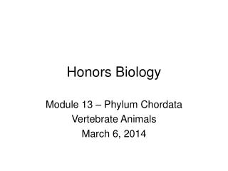
Bacteria of the “S” (smooth) strain of Streptococcus pneumoniae are pathogenic because they have a capsule that protects them from an animal’s defense system. Bacteria of the “R” (rough) strain lack a capsule and are nonpathogenic. Frederick Griffith injected mice with the two strains as shown below: CONCLUSION EXPERIMENT RESULTS Living S (control) cells Living R (control) cells Heat-killed (control) S cells Mixture of heat-killed S cells and living R cells Mouse dies Mouse healthy Mouse healthy Mouse dies Living S cells are found in blood sample. Griffith concluded that the living R bacteria had been transformed into pathogenic S bacteria by an unknown, heritable substance from the dead S cells. Can the Genetic Trait of Pathogenicity Be Transferred between Bacteria?
2. Avery • - In 1944 Avery isolated DNA, proteins, and lipids from the heat-killed, lethal bacteria, and concluded that DNA was the transforming agent.
Phage head Tail Tail fiber DNA 100 nm Bacterial cell 3. Hershey and Chase • - In 1952 Hershey and Chase used bacteria-infecting viruses containing either radioactively label S or P to show that DNA was the genetic material. Viruses Infecting a Bacterial Cell
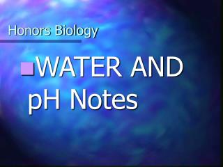
EXPERIMENT In their famous 1952 experiment, Alfred Hershey and Martha Chase used radioactive sulfur and phosphorus to trace the fates of the protein and DNA, respectively, of T2 phages that infected bacterial cells. 3 2 Agitated in a blender to separate phages outside the bacteria from the bacterial cells. Centrifuged the mixture so that bacteria formed a pellet at the bottom of the test tube. 4 1 Measured the radioactivity in the pellet and the liquid Mixed radioactively labeled phages with bacteria. The phages infected the bacterial cells. Radioactive protein Empty protein shell Radioactivity (phage protein) in liquid Phage Bacterial cell DNA Batch 1: Phages were grown with radioactive sulfur (35S), which was incorporated into phage protein (pink). Phage DNA Centrifuge Radioactive DNA Pellet (bacterial cells and contents) Batch 2: Phages were grown with radioactive phosphorus (32P), which was incorporated into phage DNA (blue). Centrifuge Radioactivity (phage DNA) in pellet Pellet Is DNA or Protein the Genetic Material of Phage T2?
Phage proteins remained outside the bacterial cells during infection, while phage DNA entered the cells. When cultured, bacterial cells with radioactive phage DNA released new phages with some radioactive phosphorus. RESULTS CONCLUSION Hershey and Chase concluded that DNA, not protein, functions as the T2 phage’s genetic material.
B. DNA Structure • - The structure of DNA was discovered by James Watson and Francis Crick in 1953. Watson and Crick with Their DNA Model
Rosalind Franklin Franklin’s X-ray diffraction Photograph of DNA • - Rosalind Franklin used X-Ray diffraction to discover the shape and dimensions of DNA.
1. Components of DNA(3 Main Parts) • c. Bases a. Sugar (Deoxyribose) b. Phosphate Deoxyribonucleic Acid
c. Bases • (1) Adenine (A) Guanine (G) • (2) Cytosine (C) Thymine (T)
2. Nucleotide: • - a subunit of a nucleic acid containing a sugar, a phosphate, and a base
3. DNA Shape: • - double helix • a. backbone - sugars and phosphates • b. paired bases form on the inside • c. Base Pairing Rule: A : T , C : G
The Watson-Crick Model of DNA Structure Link to an interview with James Watson (1:42)
II. Replication of DNA: • - process by which DNA makes an exact copy of itself • - occurs before mitosis during interphase
A A A T A T T T T G C C C G G G G C A T T T A A A A T A A A T A T T T T T G G G C G C C C C A A G C C A T T T A A C G G A. Semiconservative Replication • - DNA strands separate and serve as templates for rebuilding the other half.
DNA Replication Parental DNA double helix FreeNucleotides New double helix with 1 old &1 new strand
1. Unwinding • - DNA helicase unwinds and unzips DNA molecule. • - RNAprimaseaddsa short segment (RNA primer) on each DNA strand.
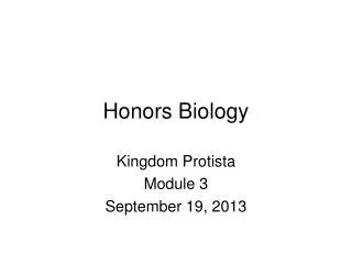
Overall direction of replication Lagging strand Leading strand Origin of replication Helicase unwinds the parental double helix. 1 Molecules of single- strand binding protein stabilize the unwound template strands. 2 The leading strand is synthesized continuously in the 5 3 direction by DNA pol III. 3 Leading strand Lagging strand OVERVIEW DNA pol III Leading strand 5 Replication fork DNA ligase DNA pol I 3 Primase 2 Parental DNA Lagging strand DNA pol III 1 Primer 3 Primase begins synthesis of RNA primer for fifth Okazaki fragment. 4 3 5 4 5 6 DNA pol III is completing synthesis of the fourth fragment, when it reaches the RNA primer on the third fragment, it will dissociate, move to the replication fork, and add DNA nucleotides to the 3 endof the fifth fragment primer. 7 DNA ligase bonds the 3 end of the second fragment to the 5 end of the first fragment. DNA pol I removes the primer from the 5 end of the second fragment, replacing it with DNA nucleotides that it adds one by one to the 3’ end of the third fragment. The replacement of the last RNA nucleotide with DNA leaves the sugar- phosphate backbone with a free 3 end. DNA Replication
2. Base Pairing • - DNA polymerase adds nucleotides with the complementary base on the 3’ end of each strand. • - The leading strand is synthesized continuously by adding nucleotides to 3’ end. • - The lagging strand is synthesized from Okazaki fragments which are later connected by DNA ligase.
3. Joining • - RNA primers are replaced by DNA nucleotides • - DNA ligase connects thefragments together. Link to DNA Replication Video Clip (2:19)
Overall direction of replication Lagging strand Leading strand Helicase unwinds the parental double helix. Origin of replication 1 Molecules of single- strand binding protein stabilize the unwound template strands. The leading strand is synthesized continuously in the 5 3 direction by DNA pol III. 2 3 Leading strand Lagging strand OVERVIEW DNA pol III Leading strand 5 Replication fork DNA ligase DNA pol I 3 Primase 2 Parental DNA Lagging strand DNA pol III 1 Primer 3 Primase begins synthesis of RNA primer for fifth Okazaki fragment. 4 3 5 4 DNA pol III is completing synthesis of the fourth fragment, when it reaches the RNA primer on the third fragment, it will dissociate, move to the replication fork, and add DNA nucleotides to the 3 endof the fifth fragment primer. DNA pol I removes the primer from the 5 end of the second fragment, replacing it with DNA nucleotides that it adds one by one to the 3’ end of the third fragment. The replacement of the last RNA nucleotide with DNA leaves the sugar- phosphate backbone with a free 3 end. DNA ligase bonds the 3 end of the second fragment to the 5 end of the first fragment. 5 6 7 A Summary of DNA Replication
1 µm Telomeres
B. DNA Replication in Prokaryotes • - Prokaryotic chromosomes made of one circular DNA strand without proteins. • - DNA replication begins at a single origin of replication and occurs very quickly.
Origins of Replication in Eukaryotes Origin of replication Parental (template) strand 0.25 µm Daughter (new) strand 1 Replication begins at specific sites where the two parental strands separate and form replication bubbles. Replication fork Bubble 2 The bubbles expand laterally, as DNA replication proceeds in both directions. 3 Eventually, the replication bubbles fuse, and synthesis of the daughter strands is complete. Two daughter DNA molecules (a) In eukaryotes, DNA replication begins at many sites along the giant DNA molecule of each chromosome. (b) In this micrograph, three replication bubbles are visible along the DNA of a cultured Chinese hamster cell (TEM).
Structure of a Eukaryotic Chromosome C. Chromosome Structure • - Chromosomes of eukaryotes are made of DNA and proteins forming bead-shaped nucleosomes which are coiled and folded to form chromatin.
C. Chromosome Structure • - Chromosomes of eukaryotes are made of DNA and proteins forming bead-shaped nucleosomes which are coiled and folded to form chromatin. Link to DNA Packaging Video Clip (1:43)
III. DNA, RNA, and Protein • A. ‘Central Dogma’ • - DNA codes for RNA which guides protein synthesis
1. Gene • - a specific sequence of bases in DNA that determines the sequence of amino acids in a protein
2. Proteins • - very complex structure • - 3 basic shapes: helix, pleated sheet, globular • - proteins contain between 50 - 2000 amino acids
Primary(Amino Acid Sequence) Illustration of Protein Structure Tertiary(Bending) Quaternary(Layering) Secondary(Helix)
Horn Hair Spiderweb Structural Proteins
HairStructure Hair Cell Single hair Microfibril Protofibril disulfide bridges |S|S| |S|S| Hydrogen bonds
StraightHair PermanentWave NaturallyCurlyHair |S|S| |S|S| |S|S| |S|S| |S|S| |S|S| |S|S| |S|S| |S|S| |S|S| |S|S| |S|S| |S|S| |S|S| Curling of Hair
B. RNA Structure: • - Nucleic acid that makes protein Ribonucleic Acid
B. RNA Structure: • DNARNA • Shape double helixsingle helix • Sugar deoxyriboseribose • Base thymineuracil • Size very largesmaller • Location nucleuscytoplasm • Function - stores genetic- makes • infoprotein • - replication • - makes RNA
Original DNA C. Transcription: • - the copying of a genetic message from DNA to RNA
C. Transcription: • - the copying of a genetic message from DNA to RNA DNA base pairs separate
C. Transcription: • - the copying of a genetic message from DNA to RNA DNA half ‘transcribes’ RNA
C. Transcription: • - the copying of a genetic message from DNA to RNA Link to Transcription Video Clip (1:54) RNA released to make protein
U U U U U C A A A A A G G G G G G G 1 2 Three Types of RNA mRNA codons Largesubunit Ribosomecontains rRNA tRNA docking sites Smallsubunit Met Amino acid tRNA anticodon
D. Messenger RNA (mRNA): • - carries the information for making a protein from DNA to the ribosomes • - acts as a template (pattern) • - contains codons: • triplets of bases that code for a particular amino acid
- Start Codon: • (AUG) - marks the start of a polypeptide • - Stop Codon: • (UAA, UAG, UGA) - marks the end
E. Transfer RNA (tRNA): • - carries amino acid to specific place on mRNA • - contains Anticodon: • triplet of bases complimentary to mRNA codon
F. Ribosomal RNA (rRNA): • - transcribed in nucleus and combined with protein into ribosomes (site of protein synthesis)
IV. Translation: • - protein synthesis • - decoding the "message" of mRNA into a protein Link to Translation Video Clip (2:05)