Download
1 / 43
1.3k likes | 4.38k Views
Introduction to CNS pharmacology. Dr. MUSTAFA A. ALSHAGGA CUCMS – Faculty of Pharmacy. Objectives. You should able to: Describe general anatomy of CNS Describe the neuron parts Understand the signals conduction Know the type of channels & receptors

E N D
Introduction to CNS pharmacology Dr. MUSTAFA A. ALSHAGGA CUCMS – Faculty of Pharmacy
Objectives • You should able to: • Describe general anatomy of CNS • Describe the neuron parts • Understand the signals conduction • Know the type of channels & receptors • Describe the steps of synaptic transmission • Describe the CNS drugs mechanism of action • Classify CNS drugs
PSYCHOSIS • A group of mental disorders (schizophrenia, depression) which are consider endogenous in origin • Delusions • Hallucinations
Neurosis • Anxiety and phobias which is regarded as abnormal reaction to external circumstances • Sweating • Tremor • Palpitation • Screaming • Panic reaction
CNS drugs: A challenging problem • Special significance tomankind • Clinical indication • Sense of wellbeing (HIGH) • Complexity of CNS • Cellular level not directly related to functional & behavioral level • Poor understanding of disease
Methods for the study of CNS Pharmacology • Microelectodes-IC recording • Patch clamp technique-Electrophysiology • Histochemical • Immunological NT & enzyme system • Radioisotopes • Molecular cloning- Receptors & enzymes • Knock-out mice
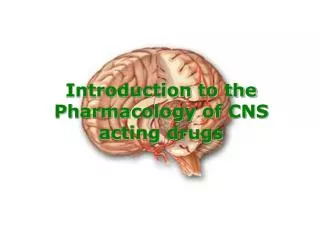
General organization of CNS • Central Nervous System • The brain + the spinal cord • The center of integration and control • Peripheral Nervous System • The nervous system outside of the brain and spinal cord • Consists of: • 31 Spinal nerves • Carry info to and from the spinal cord • 12 Cranial nerves • Carry info to and from the brain
Central Nervous System Peripheral Nervous System
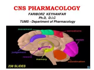
The brain Cerebral hemi. Diencephalon • Brainstem • Midbrain • Pons • Medulla oblongata • Cerebellum • Cerebrum • Cerebral hemispheres • Diencephalon • Thalamus • Hypothalamus Brainstem Cerebellum
Peripheral Nervous System • Responsible for communication btwn the CNS and the rest of the body. • Can be divided into: • Sensory Division • Afferent division • Conducts impulses from receptors to the CNS • Informs the CNS of the state of the body interior and exterior • Sensory nerve fibers can be somatic (from skin, skeletal muscles or joints) or visceral (from organs w/i the ventral body cavity) • Motor Division • Efferent division • Conducts impulses from CNS to effectors (muscles/glands) • Motor nerve fibers
Motor Efferent Division • Can be divided further: • Somatic nervous system • VOLUNTARY (generally) • Somatic nerve fibers that conduct impulses from the CNS to skeletal muscles • Autonomic nervous system • INVOLUNTARY (generally) • Conducts impulses from the CNS to smooth muscle, cardiac muscle, and glands.
Autonomic Nervous System • Can be divided into: • Sympathetic Nervous System • “Fight or Flight” • Parasympathetic Nervous System • “Rest and Digest” These 2 systems are antagonistic. Typically, we balance these 2 to keep ourselves in a state of dynamic balance.
Cells forming CNS • Neuron • Neuroglia • Astrocytes • Microglia • Ependymal cells • Oligodendrocytes
Most neurons have a single axon – a long (up to 1m) process designed to convey info away from the cell body. • Originates from a special region of the cell body called the axon hillock. • Transmit APs from the soma toward the end of the axon where they cause NT release. • Often branch sparsely, forming collaterals. • Each collateral may split into telodendria which end in a synaptic knob, which contains synaptic vesicles – membranous bags of NTs.
Ion channels & NT receptors • Voltage-gated channels • Respond to AP • Fast action potentials • Ligand-gated channels (Inotropic) • opened by binding to NT • few to tens of milliseconds • G-protein coupled receptors (Metabotropic) • Modulate voltage-gated channels • Tens of milliseconds to minutes • Direct opening voltage-channel or indirectly through SM
Chemical v/s Electrical Synapses Chemical Synapses Slow unidirectional transmission Presynaptic vesicles, active zones, postsynaptic receptors Chemical neurotransmitters Complex amplifying excitatory/ inhibitory signals Synaptic cleft Electrical Synapses Rapid bidirectional transmission Gap junctions Ion current Electrotonic transmission Cytoplasmic continuity
Action Potentials • If stimulus reaches threshold, Na+ channels open and Na+ influx ensues, depolarizing the cell and causing the AP to increase. This is the rising phase of an AP. • Eventually, the Na+ channel will have inactivated and the K+ channels will be open. Now, K+ effluxes and repolarization occurs. This is the falling phase
Chemical Signals (Synapse) • One neuron will transmit info to another neuron or to a muscle or gland cell by releasing chemicals called neurotransmitters. • The site of this chemical interplay is known as the synapse. • An axon terminal (synaptic knob) will abut another cell, a neuron, muscle fiber, or gland cell. • This is the site of transduction – the conversion of an electrical signal into a chemical signal.
Synaptic Transmission • An AP reaches the axon terminal of the presynaptic cell and causes V-gated Ca2+ channels to open. • Ca2+ rushes in, binds to regulatory proteins & initiates NT exocytosis. • NTs diffuse across the synaptic cleft and then bind to receptors on the postsynaptic membrane and initiate some sort of response on the postsynaptic cell.
EPSPs & IPSPs • Typically, a single synaptic interaction will not create a graded depolarization strong enough to migrate to the axon hillock and induce the firing of an AP. • However, a graded depolarization will bring the neuronal VM closer to threshold. Thus, it’s often referred to as an excitatory postsynaptic potential or EPSP. • Graded hyperpolarizations bring the neuronal VM farther away from threshold and thus are referred to as inhibitory postsynaptic potentials or IPSPs.
Summation • One EPSP is usually not strong enough to cause an AP. • However, EPSPs may be summed. • Temporal summation • The same presynaptic neuron stimulates the postsynaptic neuron multiple times in a brief period. The depolarization resulting from the combination of all the EPSPs may be able to cause an AP. • Spatial summation • Multiple neurons all stimulate a postsynaptic neuron resulting in a combination of EPSPs which may yield an AP
CNS DRUGS CLASSIFICATION • Chemical structure • Benzodiazepines, Butyrophenones • Pharmacological • MAO inhibitors, SSRI, Hallucinogens?? • Clinical use • Antidepressants, Antipsychotic agents, Amphetamines ??
WHO Classification 1. Anxiolytic sedatives (hypnotics, sedatives, minor tranquillizers) “Drugs that cause sleep and reduce anxiety” Examples Barbiturates, benzodiazepines and ethanol
WHO Classification 2. Antipsychotic drugs (neuroleptics, major tranquillizers, anti-schizophrenia “Drugs that are effective in relieving symptoms of schizophrenic illness” Examples Phenothiazines and butyrophenones
WHO Classification 3. Antidepressant drugs (thymoleptics) “ Drugs that alleviate the symptoms of depressive illness” Examples TCA, MAOI, SSRI
WHO Classification 4. Psychomotor stimulants (Psychostimulants) “Drugs that can cause wakefulness and euphoria” Examples Amphetamines, Cocaine, and caffeine
WHO Classification 5. Hallucinogenic drugs (psychodysleptics, psychotomimetics) “Drugs that cause disturbance of perception and of behavior in ways that cannot be simply characterized as sedative or stimulant effects” Examples Lysergic acid diethylamide (LSD), mescaline and phencyclidine
Others • Analgesics (opiate) • Katamine (anesthetics) • Nootropics drugs (Piracetam)
Home message • Pharmacology is easy • Understanding of CNS physiology and anatomy enhances understanding of drugs action • Pharmacology Range and Dale is better for further reading also read your Katzung
Neurotransmitters Lecture on 16/08/07 ?! This is just the beginning…