Download
1 / 62
630 likes | 859 Views
Introduction to the Pharmacology of CNS acting drugs. CNS acting drugs are of major therapeutic and clinical importance. They can produce diverse physiological and psychological effects such as: Induction of Anesthesia Relief of Pain Prevention of Epileptic seizures

E N D
CNS acting drugs are of major therapeutic and clinical importance. • They can produce diverse physiological and psychological effects such as: • Induction of Anesthesia • Relief of Pain • Prevention of Epileptic seizures • Reduction of Anxiety • Treatment of Parkinsonism • Treatment of Alzheimer's disease • Treatment of Depression • Centrally acting drugs also include drugs that are administered without medical intervention like tea, coffee, nicotine, and opiates.
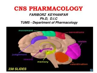
Structure of the CNS The CNS is a highly complex tissue that controls all of the body activities and serves as a processing center that links the body to the outside world. It is an assembly of interrelated “parts” and “systems” that regulate their own and each other’s activity.
Structure of the CNS • 1- Brain 2- Spinal cord • The brain is formed of 3 main parts: • I. The forebrain • the cerebrum • the thalamus • the hypothalamus • II. The midbrain • III. The hindbrain • the cerebellum • the pons • the medulla oblongata
Different Parts of the CNS & their functions The cerebrum (cerebral hemispheres): It constitutes the largest division of the brain. The outer layer of the cerebrum is known as the “cerebral cortex”.
The cerebral cortex is divided into different functional areas: • Motorareas (voluntary movements) • Sensoryareas (sensation) • Associationareas (higher mental activities • as consciousness, memory, and behavior).
Deep in the cerebral hemispheres are located the “basal ganglia” which include the “corpus striatum” & “substantia nigra”. The basal ganglia play an important role in the control of “motor” activities.
The thalamus: It functions as a sensory integrating center for well-being and malaise. It receives the sensory impulses from all parts of the body and relays them to specific areas of the cerebral cortex.
The hypothalamus: It serves as a control center for the entire autonomic nervous system. It regulates blood pressure, body temperature, water balance, metabolism, and secretions of the anterior pituitary gland.
The mid-brain: It serves as a “bridge” area which connects the cerebrum to the cerebellum and pons. It is concerned with “motor coordination”.
The cerebellum: It plays an important role in maintaining the appropriate bodyposture & equilibrium. The pons: It bridges the cerebellum to the medulla oblongata. The “locus ceruleus” is one of the important areas of the pons.
The medulla oblongata: • It serves as an organ of conduction for the passage of impulses between the brain and spinal cord. • It contains important centers: • cardioinhibitory • vasomotor • respiratory • vomiting (chemoreceptor trigger zone, CTZ).
The spinal cord: It is a cylindrical mass of nerve cells that extends from the end of the medulla oblongata to the lower lumbar vertebrae. Impulses flow from and to the brain through descending and ascending tracts of the spinal cord.
Different Systems of the CNS & their functions These systems are pathways formed of specific parts of the brain and the neurons connecting them. They include: 1. The pyramidal system 2. The extrapyramidal system 3. The limbic system 4. The reticular formation 5. The tuberohypophyseal system
The pyramidal system: It originates from the motor area of the cerebral cortex and passes through the spinal cord, therefore it is also known as the “corticospinal tract”. It is responsible for the regulation of the finevoluntarymovements.
The extrapyramidal system: It also controls the motor function but involves areas other than the corticospinal tract. It is involved in the regulation of grossvoluntary movements, thus it complements the function of the pyramidal system. The “basal ganglia” constitute an essential part of this system. Degenerative changes in the pathway running from the “substantia nigra” to the “corpus striatum” (or nigrostriatal pathway) may cause tremors and muscle rigidity characteristic of “Parkinson’s disease”.
The limbic system: The major parts of this system are: the hypothalamus, the basal ganglia, the hippocampus (responsible for short term memory), and some cortical areas. The limbic system is involved in the control of “behavior” & “emotions”.
The reticular formation: It is composed of interlacing fibers and nerve cells that run in all directions beginning from the upper part of the spinal cord and extending upwards. It is important in the control of “consciousness” and “wakefulness”. The tuberohypophyseal system: It is a group of short neurons running from the hypothalamus to the hypophysis (pituitary gland) regulating its secretions.
Cells of the Nervous System 1- Neurons (Nerve Cells):function units of the nervous system by conducting nerve impulses, highly specialized and amitotic. Each has a cell body (soma), one or more dendrites, and a single axon. • Cell Body:it has a nucleus with at least one nucleolus and many of the typical cytoplasmic organelles, but lacks centrioles for cell division. • Dendrites:Dendrites and axons are cytoplasmic extensions (or processes), that project from the cell body. They are sometimes referred to as fibers. Dendrites (afferent processes) increase their surface area to receive signals from other neurons, and transmit impulses to the neuron cell body. • Axon: There is only one axon (efferent process) that projects from each cell body. It carries impulses away from the cell body.
Cells of the Nervous System 2- Glial cells: do not conduct nerve impulses, but support, nourish, and protect the neurons. They are mitotic, and far more numerous than neurons. • Astrocyte:A glial cell that provides support for neurons of the CNS, provides nutrients regulates the chemical composition of the extracellular fluid.
Cells of the Nervous System • Oligodendrocyte:A type of glial cell in the CNS that forms myelin sheaths.
Cells of the Nervous System • Microglia:The smallest glial cells; act as phagocytes (cleaning up debris) and protect the brain from invading microorganisms.
Cells of the Nervous System • Schwann cell:A cell in the PNS that is wrapped around a myelinated axon, providing one segment of its myelin sheath.
Types of Neurons (Function) • There are 3 general types of neurons (nerve cells): 1- Sensory (Afferent ) neuron:A neuron that detects changes in the external or internal environment and sends information about these changes to the CNS. (e.g: rods and cones, touch receptors). They usually have long dendrites and relatively short axons. 2- Motor (Efferent) neuron:A neuron located within the CNS that controls the contraction of a muscle or the secretion of a gland. They usually have short dendrites and long axons. 2- Interneuron or association neurons:A neuron located entirely within the CNS in which they form the connecting link between the afferent and efferent neurons. They have short dendrites and may have either a short or long axon.
Types of Neurons (Morphology) 1- Unipolar neuron: A neuron with one axon attached to its soma. • the axon divides, with one branch receiving sensory information and sending the information into the CNS.
2- Bipolar neuron: A neuron with one axon and one dendrite attached to its soma.
3- Multipolar neuron: A neuron with one axon and many dendrites
Neuron Basic Structure (How brain cells communicate) • Synapse:A junction between the terminal button of an axon and the membrane of another neuron • Terminal button(or bouton): The bud at the end of a branch of an axon; forms synapses with another neuron; sends information to that neuron. • Neurotransmitter: A chemical that is released by a terminal button; has an excitatory or inhibitory effect on another neuron.
Different types of Synapses1- Axo-denrdritic 2- Axo-axonal 3- Axo-somatic
Chemical transmission in the CNS The CNS controls the main functions of the body through the action endogenous chemical substances known as “neurotransmitters”. These neurotransmitters are stored in and secreted by neurons to “transmit” information to the postsynaptic sites producing either excitatory or inhibitory responses. Most centrally acting drugs exert their actions at the synaptic junctions by either affecting neurotransmitter synthesis, release, uptake, or by exerting direct agonist or antagonist action on postsynaptic sites.
Neurotransmitters can be classified into: 1. Biogenic amines: ACh, NA, DA, 5-HT, Histamine 2. Amino acids: Excitatory (glutamate & asparate) Inhibitory (GABA & glycine) 3. Others: Adenosine, melatonin
Acetylcholine • - Acetylcholine is widely distributed in major parts of the CNS. • Cholinergic neurons occur particularly in the motor areas of the cerebral cortex, the corpus striatum, the hippocampus, and the spinal cord. • - Both types of Cholinergic receptors (nicotinic & muscarinic) occur in the CNS. • Muscarinic receptors (predominantly M1) are much more abundant than nicotinic receptors and mediate many of the behavioral effects of acetylcholine in cholinergic pathways. • Muscarinic receptors are located presynaptically and upon stimulation by agonist inhibits Ach release, while muscarinic antagonists increase Ach release.
Acetylcholine in the brain plays a role in 1- Arousal 2- Learning 3- Motor control 4- Short term memory - Loss of cholinergic neurons in thehippocampus(memory site) is associated with “Alzheimer’s disease (The most common neurodegenerative disease causing progressive irreversible dementia associated with old age.
Cholinergic drugs andAlzheimer's disease • Reversible cholinesterase inhibitors:Tacrine, Donepezil and Galantamine. • Pseudo-reversible cholinesterase inhibitors:Physostigmine, Eptastigmine and Rivastigmine. • Irreversible cholinesterase inhibitors: Metrifonate
- Hyperactivity of cholinergic neurons in the corpus striatum leads to “Parkinson’s disease”. - Parkinsonism, is a progressive neurodegenerative brain disorder that was first described by Dr. James Parkinson in 1817. - The disease occurs above the age of 65 and affects the "extrapyramidal system" at the level of the "corpus striatum" & "substantianigra"
In the basal ganglia, the 2 neurotransmitters "dopamine" & "acetylcholine" co-exist in a normal balance, where dopaminergic neurons exert an inhibitory effect on excitatory cholinergic neurons. DA ACh In Parkinson’s disease, the "nigrostriatal dopaminergic neurons" are degenerated (damaged) while cholinergic neurons are intact, resulting in a marked decrease in dopamine content (dopamine deficiency syndrome) and a relative predominance of cholinergic activity. Therefore, the strategy for treatment of Parkinsonism is to restore the normal balance between dopaminergic & cholinergic tones in the basal ganglia.
Treatment of Parkinsonism • • D2 • • • • • D2 • • • Tyrosine DOPA DA • • • • • • • D2 • • • MAO-B • • D2 • Dopaminergic Nerve terminal Postsynaptic receptors
Drugs used to treat Parkinsonism do not stop the progression of the disease but they can only ameliorate the symptoms. Parkinsonism could be managed by: A.Enhancing dopaminergic activity using: • Reducing cholinergic activity by the use of anticholinergic drugs (benzatropine) • Neurotransplantation (which is still in experimental phase). • Physical therapy & regular exercise. • drugs that replace dopamine (levodopa) • drugs that release dopamine from its stores (amantadine) • drugs that prolong the action of dopamine by preventing its metabolism (seligiline) • drugs that mimic the action of dopamine (dopaminergic agonists)
Noradrenaline Noradrenergic neurons arise mainly from the “locus ceruleus” (in the pons) and the reticular formation and project diffusely to many areas of the brain. As in the periphery, the same types of adrenergic receptors are described in the CNS. Noradrenaline plays an important role in the regulation of both “arousal” and “mood”.
Noradrenaline plays a role in 1- Mood: Deficiency of noradrenaline in certain parts of the brain is thought to be the underlying cause of “depression”. 2- Arousal: Increased release of noradrenaline in the brain is responsible for wakefulness and alertness. 3- Blood pressure regulation: In addition, noradrenergic synapses form a part of the baroreceptor reflex pathway that regulates blood pressure.
Dopamine Dopamine acts as both a “precursor” for noradrenaline by the action of the enzyme “dopamine β-hydroxylase” in noradrenergic neurons, as well as a “neurotransmitter” in dopaminergic neurons which lack this enzyme. Dopamine receptors are of 2 types: • D1-type (including D1 & D5 subtypes) • D2-type (including D2, D3, & D4 subtypes) Both types are G-protein-coupled receptors that involve “adenylate cyclase/cAMP” as a signal transduction mechanism (D1-receptors activate, while D2-receptors inhibit adenylate cyclase).
Dopamine is the major neurotransmitter of the 3 following systems: • The nigrostriatal system • The mesolimbic system • The tuberohypophyseal system
Dopamine plays a role in 1- Motor activity: In the nigrostriatal system, dopamine is involved in the control of motor function. Deficiency of dopamine in this system causes “Parkinson’s disease”. 2- Behavioural effects: In the mesolimbic system, dopamine is involved in the control of behavior and emotion. Increased dopaminergic activity in this system is believed to cause “schizophrenia”.
3- Anterior pituitary function: In the tuberohypophysealsystem, dopamine inhibits the secretion of prolactin. Bromocriptine (dopaminergic agonist) is used clinically for this purpose. Dopamine stimulates the secretion of growth hormone from the pituitary gland in normal subjects, but inhibits its release in acromegaly, a condition in which bromocriptine is useful therapeutically if given before excessive growth has taken
4- Vomiting: • Dopamine has a role in the production of “nausea & vomiting” by stimulating dopamine receptors in the chemoreceptor trigger zone (CTZ) in the medulla oblongata. • Therefore, dopaminergic antagonists as Metoclopramide and phenothiazines have antiemetic properties. • Dopaminergic agonists as Bromocriptine and levodopa cause nausea and vomiting as side-effects.
5-Hydroxytryptamine (5-HT) 5-HT is an important CNS transmitter although the brain accounts for only 1% of its total body content. 5-HT-containing neurons arise from nuclei in the pons and medulla known as the “raphenuclei” and project diffusely to many areas of the brain similar to noradrenergic neurons. Formation and release of 5-HT is very similar to NA. Many drugs as TCA that inhibit NA uptake, also inhibit 5-HT uptake.
5-HT receptors • 5-HT receptors in the CNS include 1- 5-HT1 receptors: located presynaptically and act to inhibit transmitter release. They includes 5 subtypes(5-HT1A,5-HT1B, 5-HT1C,5-HT1D, 5-HT1E) 2- 5-HT2 receptors: located post-synaptically and may exhibit excitatory or inhibitory functions. They includes 3 subtypes (5-HT2A, 5-HT2B, 5-HT2C) 3- 5-HT3 receptors. • 5-HT pathways in the CNS are involved in behavioral changes, mood, hallucinations, sleep, wakefulness, and control of sensory transmission.
5-HT plays a role in 1- Mood: increasing 5-HT levels by giving tryptophan is useful in depressive states. 2- Sensory transmission: 5-HT inhibits transmission of pain impulses in the spinal cord and brain and enhances morphine analgesia. 3- Temperature control: the hypothalamus is rich in 5-HT containing neurons. Experimentally, injection of 5-HT in that region causes a rise in body temperature, while exposure to cold climates increases 5HT release. 4- Anterior pituitary function: The secretion of the hypothalamic releasing factors for certain anterior pituitary hormones as gonadotrophins is influenced by 5-HT containing neurons in the hypothalamus. 5- Vomiting: 5-HT3 blocker (ondansetrone) is used as anti-emetic.