Download
1 / 7
70 likes | 72 Views
A process that examines inside organs and tissues using high-energy sound waves. On a computer screen, the echoes created by the sound waves create images of the tissues and organs (sonogram). Read about its risks, preparation and results. Click on below link: https://www.hillregionalhospital.com/blogs/what-is-an-ultrasound/<br><br><br><br><br>

E N D
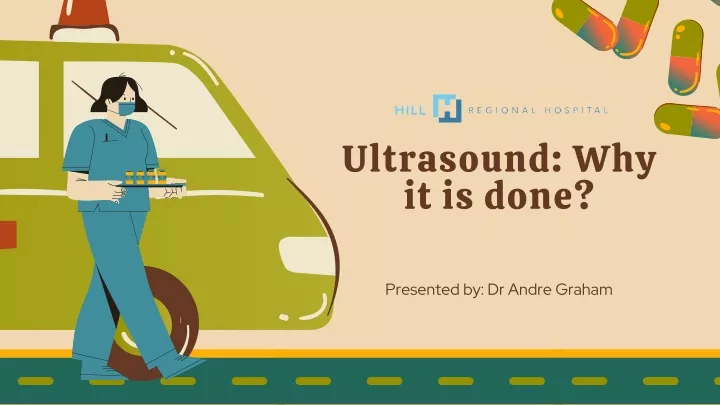
Ultrasound: Why it is done? Presented by: Dr Andre Graham
Topic Outline WHY IT IS DONE? RISKS PROCEDURE RESULTS
Why it is done? Ultrasound is used for a number of reasons, including to: Observe the uterus and ovaries during pregnancy and monitor the developing baby's health Diagnose gallbladder disease Evaluate blood flow Guide a needle for biopsy or tumor treatment Examine a breast lump Check the thyroid gland Find genital and prostate problems Assess joint inflammation (synovitis) Evaluate metabolic bone disease
Ultrasound Risks Diagnostic ultrasound is a low-risk procedure that uses low-power sound waves. There aren’t any known risks. Ultrasound is a valuable tool, but it has restrictions. Sound waves do not travel well through air or bone, so ultrasound is not effective at imaging body parts that have gas in them or are hidden by bone, like the lungs or head. Ultrasound might also be unable to see objects that are located very deep in the human body. To view these areas, your health care provider might order other imaging tests, such as CT or MRI scans or X-rays.
Ultrasound Preparation For some scans, such as a gallbladder ultrasound, your care provider might ask that you not eat or drink for a certain period of time before the examination. Others, such as a pelvic ultrasound, might require a full bladder. Your doctor will let you know how much water you need to drink before the examination. Do not urinate until the examination is done. Young children might need additional preparation. When scheduling an ultrasound for yourself or your child, ask your doctor if there are any specific instructions you will need to follow. Wear loose clothing for your ultrasound appointment. You might be asked to remove jewelry during your ultrasound, so it is a good idea to leave any valuables at home.
Ultrasound Results When your examination is complete, a doctor trained to interpret imaging studies (radiologist) analyzes the pictures and sends a report to your doctor. Your doctor will let you know about the results. You should be able to get back to normal activities immediately after an ultrasound. When your examination is complete, a doctor trained to interpret imaging studies (radiologist) analyzes the pictures and sends a report to your doctor. Your doctor will let you know about the results. You should be able to get back to normal activities immediately after an ultrasound.
Contact Address: 101 Circle Drive Hillsboro, TX 76645 Contact: 254-580-8500. Website: https://www.hillregionalhospital.com/blogs/what-is-an-ultrasound/