Download
1 / 29
451 likes | 2.33k Views
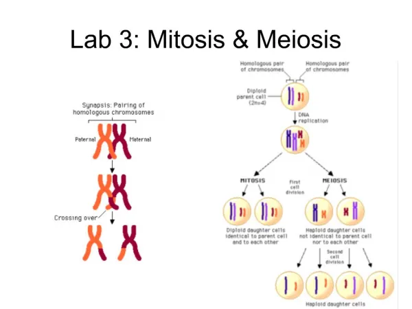
BIO1140 Lab 5 Meiosis. Objectives. Identify selected meiotic phases and their characteristics Microsporogenesis in Lilium Meiosis in Ascaris . Identify various stages during micro- sporogenesis . Study spermatogenesis in the rat testis and oogenesis in the rabbit ovary.

E N D
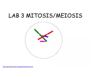
BIO1140 Lab 5 Meiosis BIO1140 Introduction to Cell Biology
Objectives • Identify selected meiotic phases and their characteristics • Microsporogenesis in Lilium • Meiosis in Ascaris. • Identify various stages during micro-sporogenesis. • Study spermatogenesis in the rat testis and oogenesis in the rabbit ovary. • Capture digital images of assigned material and draw a biological drawing based on those photos. BIO1140 Introduction to Cell Biology
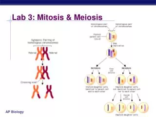
Meiosis Reductive division of cells Diploid cell (2n) Pre-mieotic interphase Duplication of chromosomes tetraploid cell (4n) Separation of homologouschromosomes First division (2n) Separation of chromatids Second division haploid cells (n)
I- Meiosis in plants Lily microsporogenesis • Formation of male gametes (pollen) in Anther • Identify stages and structures using prepared slides • visit also website: http://www.iasprr.org/old/iasprr-pix/lily/male.shtml BIO1140 Introduction to Cell Biology
Meiosis in plants pa f anther pc e tapetum Meiosis in Lillium anther (early prophase): e: epidermis; f: filament; pa: parenchyme; pc: pollen cell precursor (prophase) pollen sacs (microsporangia)
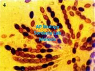
II- Meiosis in Animals Meiosis in Ascaris BIO1140 Introduction to Cell Biology
Selected stages Sperm entrance Primary oocyte arrested in prophase 1 (no shell) Penetration of the spermatozoid (sp) triggers the egg nucleus (n) to continue meiosis sp n BIO1140 Introduction to Cell Biology
Selected stages Metaphase I Presence of shell (s) Chromosomes lined up at equator (c) No polar body Male pronucleus visible (p) at center of cell p s c BIO1140 Introduction to Cell Biology
Selected stages Anaphase I Homologous chromosomes separate and are moving to opposite spindle poles BIO1140 Introduction to Cell Biology
Selected stages Metaphase II Similar to metaphase I but with the presence of a polar body (pb) in the perivitelline space (ps) ps pb BIO1140 Introduction to Cell Biology
Selected stages Anaphase II Centromeres split chromatids separate then move to cell poles BIO1140 Introduction to Cell Biology
Selected stages Interphase Following telophase II, haploid male and female pronuclei (pn) in interphase prior to fusion (fertilization). Second polar body expelled (not visible here) The resulting zygote (2n) continues to divide by mitosis. pn BIO1140 Introduction to Cell Biology
Selected stages Mitotic cleavage of the embryo BIO1140 Introduction to Cell Biology
Meiosis in mammals • Gametogenesis in rat testis and rabbit ovary • You’ll be assigned one species for your lab report • Observe both species (exam material) BIO1140 Introduction to Cell Biology
Rat (Rattusnorvegicus) spermatogenesis • Occurs within the walls of the seminiferous tubules • Recognize stages • by position (immature stages near outside of tubule, mature stages near lumen). • See http://w3.ouhsc.edu/histology/for details BIO1140 Introduction to Cell Biology
sc bl ps st sg ss sp L bl: basal lamina, L: lumen of the tubule, sc: sertoli cell nucleus, sg: spermatogonia, ps: primary spermatocyte, ss: secondary spermatocyte, sp: sperm cells (cross section), st: spermatids BIO1140 Introduction to Cell Biology
Rabbit (Sylvilagusfloridanus)oogenesis Identify maturation stages of the follicles depending on position in the ovary, number of layers of follicular cells and presence of fluid-filled cavities See http://w3.ouhsc.edu/histologyfor details BIO1140 Introduction to Cell Biology
Primordial follicle Primordial follicle (pf) are located underneath the ovary epithelium (ep) Surrounded by one layer of squamous (=flat) follicular cells ic: interstitial cells pf ic ep BIO1140 Introduction to Cell Biology
Primary unilaminar follicle Located deeper in the ovary Surrounded by one layer of cuboidal follicular cells (fc) Contains a primary oocyte (po) n: oocyte pronucleus po n fc BIO1140 Introduction to Cell Biology
Growing follicle (primary multilaminar): Slightly deeper in the ovary Surrounded by several layers of follicular cells (fc) Contain primary oocyte (dash lines). n: oocyte pronucleus fc n BIO1140 Introduction to Cell Biology
Mature or Graafian Follicle Largest follicle Many layers of follicular cells (fc) One big or several fluid-filled cavities (fca) Contains secondary oocyte (so) stopped in metaphase II = mature oocyte Comes near to ovarian epithelium prior to ovulation so fc fca BIO1140 Introduction to Cell Biology
Lab5 Report: 3 pages Page 1: Title page Page 2: Annotated picture printout Page 3: Biological drawing You must take 2 pictures during the lab that you will use for your lab report: BIO1140 Introduction to Cell Biology
Rabbit ovary (Sylvilagus floridanus) • Picture 1: Global view at 10x showing follicles at various stages (at least 2 stages, such as primordial and unilaminar) plus (if possible) ovarian epithelium. • Picture 2: close-up view at 40x of picture showing a unilaminar or multilaminar follicle. BIO1140 Introduction to Cell Biology
Rat testis (Rattus norvegicus) • Picture 1 : Global view at 10x of a cross section showing several seminiferous tubules. • Picture 2: magnified view at 40x of picture 1 showing both the lumen and the periphery of one seminiferous tubule. BIO1140 Introduction to Cell Biology
Lab5 Report • Annotated picture (picture 1): use template file (download on lab website) to print picture (grayscale), then label either on computer or by hand. • Follow biological drawing instructions relative to labels and caption • Biological drawing (picture 2): read instructions in lab manual appendix. Remember: no shading, stippling, patterns, all lines have same thickness. BIO1140 Introduction to Cell Biology
Lab5 Report • Take pictures showing the required view and structures then call TA so they can check the content of your pictures and evaluate their quality • Do not leave without saving (and/or emailing) the pictures or you won’t have any material for your assignment. • Add a scale bar AND a caption to both the printout and the drawing pages BIO1140 Introduction to Cell Biology
Lab 5 Report and evaluation Available documents (lab manual and/or Lab web): • List of structures you can observe on sections - use this list to label your drawing. • Instruction file for lab5 and biological drawing. • Guide Biolabo: labeling, scale bar and caption sections of the “Biological drawings” chapter. Don’t forget: • Tech skill marks: 15% of lab5 mark (cleanliness, microscope use and pictures taken). BIO1140 Introduction to Cell Biology
Corrected lab5 reports and Final lab marks • Corrected lab5 reports will be available to pick up about 3 weeks after your lab (date and location will be announced on website). • All lab marks (including the final mark) will be posted on the website (date TBA). • Corrector office hours are posted on the contact page of website. If the corrector for your section is not listed, contact your TA. BIO1140 Introduction to Cell Biology
FINAL EXAM: SATURDAY MARCH 29TH 1PM-2:30PM • Contact coordinator today if you have a conflict with another course or exam. • Exam rooms: will be posted on Final exam page of website • Materiel covered: all Lab manual content (including appendices), lab experiments, specimens (including Lab5 structure list) and lab reports. • Format: multiple choice, short answers and identification of structures on pictures. similar to prelab quizzes. BIO1140 Introduction to Cell Biology