Download
1 / 46
720 likes | 1.78k Views
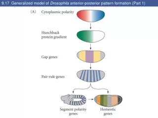
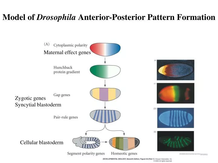
Model of Drosophila Anterior-Posterior Pattern Formation. Maternal effect genes. Zygotic genes Syncytial blastoderm. Cellular blastoderm. Homeotic selector genes Similar signal into different structures— Different interpretation—controlled by Hox genes.

E N D
Model of Drosophila Anterior-Posterior Pattern Formation Maternal effect genes Zygotic genes Syncytial blastoderm Cellular blastoderm
Homeotic selector genes Similar signal into different structures— Different interpretation—controlled by Hox genes
Homeotic transformation of the wing and haltere Homeotic genes—mutated into homeosis transformation As positional identity specifiers: Bithorax-haltere into wing
Imaginal discs and adult thoracic appendages Bithorax mutation—Ubx misexpressed T3 into T2 –anterior haltere into Anterior wing Postbithorax muation (pbx)— Regulatory region of the Ubx— Posterior of the haltere into wing
The spatial pattern of expression of genes of the bithorax complex Bithorax—Ultrabithorax –5-12 Abdominal-A—7-13 Abdominal-B—10-13 Bithorax mutant –PS 4 default state +Ubx—5,6 +Abd-A—7,8,9 +Abd-B—10 Combinatorial manner Lack Ubx—5,6 to 4 also 7-14 thorax structure in the abdomen Hox—gap, pair-rule for the first 4 hours, then polycomb (repression), and Trithorax (activation)
Segmental identity of imaginal disc Antennapedia—expressed in legs, but not in antenna If in head, antennae into legs Hth (homothorax) and Dll (distal-less)—expressed in antennae and leg In antenna: as selector to specify antenna In leg: antennapedia prevents Hth and Dll acting together Dominant antennapedia mutant (gene on)— blocks Hth and Dll in antennae disc, so leg forms No Hth, antenna into leg
Egg chamber formation A/P during oogenesis the oocyte move towards one end in contact with follicle cells. Both the oocyte and the posterior follicle cells express high levels of the E-cadherin If E-cadherin is removed, the oocyte is randomly positioned. Then the oocyte induces surrounding follicle cell to adopt posterior fate.
Specifying the Anterior-Posterior Axis of the Drosophila Embryo During Oogenesis
Specifying the Anterior-Posterior Axis of the Drosophila Embryo During Oogenesis Protein kinase A orients the microtubules
Anterior/posterior extremities Torso---receptor tyrosine kinase Ligand---trunk Terminal structure- acron., telson, most posterior abdominal segment Before fertilization ligand immobilized Small quantities—bound to torso at the poles little left to diffuse
Torso signaling Groucho: repressor Huckenbein, tailless are released from transcriptional suppression
The EGFR signal establishes the A/P and D/V axial pattern Gurken—TGFa Torpedo--- EGFR
The EGFR signal establishes the A/P and D/V axial pattern Red-actin Green-gurken protein As well as mRNA The expression of EGFR pathway target gene
The localization of Gurken RNA Cornichon, and brainiac- Modification and Transportation of the protein K10, squid localize gurken mRNA Cappuccino and spire – cytoskeleton of the oocyte
The Key determinant in D/V polarity is pipe mRNA in follicle cells Cross section
The activation of Toll windbeutel—ER protein pipe—heparansulfate 2-o-sulfotransferase (Golgi) nudel—serine protease
The dorsal-ventral pathway Perivitelline space Fig. 31-16
Toll pathway Maternal genes— Fertilization to cellular blastoderm Dorsal system—for ventral structure (mesoderm, neurogenic ectoderm) Toll gene product rescue the defect Toll mutant – dorsalized (no ventral structure) 2. Transfer wt cytoplasm into Toll mutant specify a new dorsal-ventral axis (injection site =ventral side) spatzle (ligand) fragment diffuses throughout the space
The mechanism of localization of dorsal protein to the nucleus Without Toll activation Dorsal + cactus Toll activation – tube (adaptor) and pelle (kinase) Phosphorylate cactus and promote its degradation B cell gene expression Dorsal=NF-kB Cactus=I-kB
The dorsal-ventral pathways Fig. 31-17
Dorsal nuclear gradient Activates—twist, snail (ventral) Represses—dpp, zen (dorsal) Fig. 31-19
Toll protein activation results in a gradient of intranuclear dorsal protein Spatzle is processed in the periviteline space after fertilization
Model for the subdivision of the dorso-ventral axis into different regions by the gradient in nuclear dorsal protein Zygotic genes pattern the early embryo Dorsal protein activates twist and snail represses dpp, zen, tolloid Rhomboid----neuroectoderm Repressed by snail (not most ventral) Binding sites for dorsal protein in their regulatory regions
Nuclear gradient in dorsal protein Dorsalized embryo— Dorsal protein is not in nuclei Dpp is everywhere Twist and snail are not expressed Threshold effect—integrating Function of regulatory binding sites Regulatory element =developmental switches High affinity (more dorsal region-low conc.) Low affinity (ventral side-high conc.)
Dpp protein gradient Cellularization---signal through transmembrane proteins Dpp=BMP-4(TGF-b) Dpp protein levels high, increase dorsal cells Short of gastrulation (sog) prevent the dpp spreading into neuroectoderm Sog is degraded by tolloid (most dorsal)
The TGK-b/Bmp signaling pathway • Antagonist • Proteases dpp: decapentaplegic Smad= Sma + Mad Sma-C. elegans Mad-Fly Fig. 31-24
The Wnt and BMP pathways are used in early development Fig. 31-23
Signal Pathways Induced by Cellular Surface Receptors Mol. Cell. Biol. 5th ed. 2004, Lodish et al.
The Smad-dependent pathway activated by TGF-b Type I, II receptor-Ser/Thr phosphorylation
The Smad-dependent pathway activated by TGF-b Colorectal cancer: type II receptor Pancreatic cancers: 50% Smad One component between receptor and gene regulation
De-repression of target genes in Dpp signaling Nature reviews genetics-8-663-2007
Structural and Functional Domains of Smad Family TGFb , Activin: R-Smad 2,3 BMPs: R-Smad 1, 5, 8 Common Smad4-nucleocytoplasmic shuttling, DNA binding Inhibitory Smads: I-Smad 6, 7 Cell, 95,737,1998
Smad4 shuttles between the cytosol and nucleus NLS , NES 13,216, 2003
Inhibitory Smads: I-Smad 6, 7 —recruting Smurf (ubiquitin ligase to receptor) Cell, 95,737,1998
Different internalization pathways resulted in distinct cellular effects 2005, 17:107
Models of morphogen gradient formation sharpen Fig. 31-11, 12, 13
Integration of two signal pathways at the promoter Smad2 and FAST Smad3 and c-Jun/cFos Cell,95,737, 1998 SBE: Smad binding element ARE: activin-response element TRE: TPA-response element (AP-1 binding) XBE: transcription X
The axis determining systems Fig. 31-21