Download
1 / 21
210 likes | 480 Views
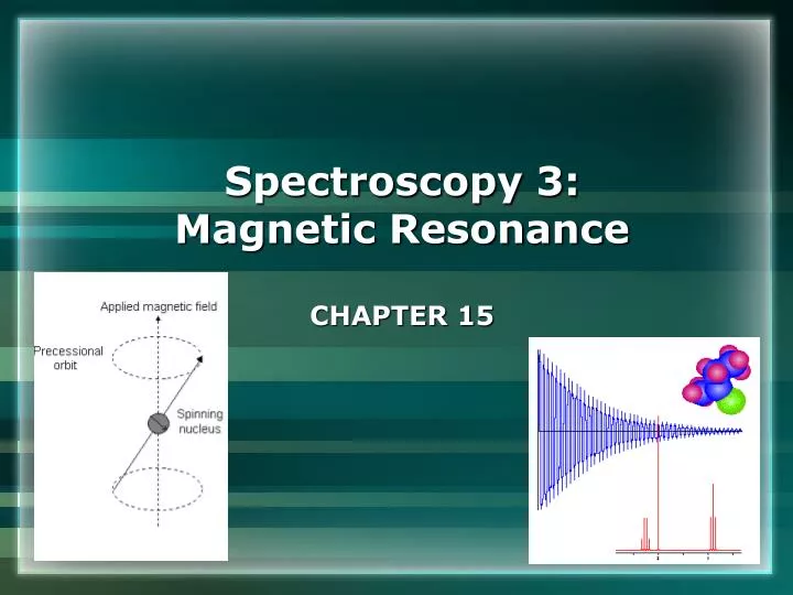
Spectroscopy 3: Magnetic Resonance CHAPTER 15. Conventional nuclear magnetic resonance Energies of nuclei in magnetic fields Typical NMR spectrometer The chemical shift (effect of nearby nuclei) Fine structure (nuclear spin-spin coupling) Pulsed techniques in FT-NMR.

E N D
Spectroscopy 3: Magnetic Resonance CHAPTER 15
Conventional nuclear magnetic resonance • Energies of nuclei in magnetic fields • Typical NMR spectrometer • The chemical shift (effect of nearby nuclei) • Fine structure (nuclear spin-spin coupling) • Pulsed techniques in FT-NMR
Fig 15.1 Interactions between ms states of an electron and an external B field precession νL ≡ the Larmor freq ms = +1/2 where γe≡ magnetogyric ratio Bo ≡ applied magnetic field ms = −1/2
Fig 15.3 Nuclear spin states of a spin-1/2 nucleus (e.g., 1H or 13C) in a magnetic field • Typically: • A 100 MHz NMR • employs a 2.35 T field • Resonance is achieved • when νradio = energy • separation between • levels = hνradio
The Chemical Shift • Nuclear magnetic moments interact with the local field • In most cases, Bloc ≠ B0 due to electronic orbital ang momentum • The Larmor frequencyνL (frequency of precession) • differs for nuclei in different environments • Resonance frequencies expressed as the chemical shift TMS
Fig 15.5(a) Range of typical chemical shifts for 1H TMS Deshielded nuclei (low field) Shielded nuclei (high field)
Fig 15.5(b) Range of typical chemical shifts for 13C TMS Shielded nuclei (high field) Deshielded nuclei (low field)
Fig 15.6 The 1H-NMR spectrum of ethanol singlet triplet quartet 3 2 1 Integrated signal
Fig 15.7 Variation of the chemical shift with electronegativity Trend due to magnetic anisotropy Trend due to electronegativity
Fig 15.9 Ring current deshields ring protons and shields substituent protons • Special case of neighboring group effect in aromatics shielded deshielded
Fig 15.6 The 1H-NMR spectrum of ethanol singlet triplet Fine structure quartet 3 2 1 Integrated signal
Fine Structure • Each magnetic nucleus may contribute to the local field of • adjacent nuclei • ∴ Resonance frequencies are modified • Strength of interaction given by the coupling constant, J (Hz) • J is independent of applied mag field, Bo
Margin pg 526 n equivalent nuclei split adjacent spin(s) into n+1 lines with intensity distribution given by Pascal’s triangle:
Fig 15.15 Origin of the 1:2:1 triplet in the proton resonance of a –CH3 species e.g., CH3CH2OH ⇆ B0 ⇉ ⇇ ⇄
Fig 15.16 Origin of the 1:3:3:1 quartet in the proton resonance of a -CH2- species B0 e.g., CH3CH2OH