Download
1 / 97
1.51k likes | 2.47k Views
MASS SPECTROSCOPY. Mass spectroscopy is a technique that determine the molecular weight and elemental compostion of a compound accurately. It recorded relative abundance and mass / charge(m/e) ratio. Relative abundance means number of different kinds of ions.

E N D
Mass spectroscopy is a technique that determine the molecular weight and elemental compostion of a compound accurately. • It recorded relative abundance and mass / charge(m/e) ratio. • Relative abundance means number of different kinds of ions.
It is used to determine the mol.weight, determine the stable isotope, examination of ionization phenomena. Free radical studies • Very small amt is needed for analysis(only picogram)
History • Wien’s determine the +ve ion could deflected the EMR in 1898. • Thomson was developed the instrument in 1905. • Aston and Dempster are furherdevolped the instrument in 1979.
Principle • Vapour form of Organic comp is bombarded with Electron beam under the pressure btwn 10-7to 10-5 mm of Hg. • The molecules are broken into highly energetic positive charge ions(molecular ion or parent ions.) • The formation is due to loss on electron from the molecule.
This radical ion can further break up into smaller ion (fragment ion or daughter ion) • This positive ions are accelerated by an electric field. • it is sorted out According to mass charge ratio by deflection in EMR. • These ions are collected by a detector and produce the mass spectrum.
Mass spectrum is a plot of m/z ratio of the ionic species present versus relative abundance. • The mol. Wt can be determined from the formation of the fragment ion and their abundance.
Compound is bombarded with a beam of electrons. It produce an ionic molecule / ionic fragments. Separation of fragments. Spectrum- mass spectrum. Place fn gps into certain areas of the molecule and see how they are interconnected.
Mass Spectrum : presentation • A graph of m/z Vs. Relative abundance. • Where, • M is the mass of the ion and z is the charge of the ion. • Relative abundance is the number of times an ion of that m/z ratio strikes the detector.
MOLECULAR ION/ PARENT PEAK • When a sample substance is bombarded with electrons of energies of 9-15 eV, the molecular ion is produced by loss of a single electron. • This will give rise to a very simple mass spectrum with essentially all of the ions appearing in one peak…
BASE PEAK • If an electron beam of energy of 70 eV is used in a mass spectrometer, the molecular ion is produced by the loss of a single electron which undergoes splitting to form many fragments. • The parent peak in the mass spectrum is called the base peak and the heights of all other peaks are measured with respect to it.
Negative ions • An electron is removed from the molecule giving a radical ion. • The molecule, giving radical anion, captures an electron. Rearrangement ions: it produced from some molecules bond breakage accompained by transfer of Hydrogen atoms are group like alkile, aryl. ….
Process of ionization • Ionization of the sample • Acceleration of ions by electric field. • Dispersion of ions according to m/e ratio • Detection of ions produce a corresponding ionic current.
Instrumentation • Sample inlet • Ion source • Ion seperator • Detector • Recorder
INSTRUMENTATION Basic components • Sample handling system • Ionisation chamber / ion source • Electrostatic accelerating system • Magnetic field • Ion separator / mass analyser • Ion collector, detector & read out system • Vacuum system
Sample handling system • M sample( gaseous state) slowly introduced in Ionisation chamber • Pressure in sample reservoir is kept greater than ionisation chamber to maintain steady flow of sample • Sample inlet systems are different for different samples • Heated Inlet system • Direct Inlet system • Gas chromatographic Inlet system
Heated Inlet system • used for gaseous & less volatile liquid samples with bp < 5000C • sample is vaporised externally & then slowly introduced into ionisation chamber with the help of vacuum locks for maintaining the vacuum • Volatile liquids are handled byFreeze out technique. The sample in sample holder tube is frozen with liquid nitrogen or dry ice & after evacuation, the tube is heated to room temp or higher to evaporate the sample into sample reservoir • Low volatile samples like sugar, acids, AA, etc. are converted into volatile dervs. • e.g. -OH to -OR, -COOH to -COOR
Direct Inlet system • Solids, nonvolatile liquids and thermally unstable compds are directly introduced into the ion source with sample probe by inserting through a vacuum lock • In the probe, there is a provision for heating as well as cooling of sample • The sample in the probe is slowly vaporised in the electron bombardment region • A fraction of mg is sufficient to record the spectrum • Non- volatile samples like steroids, polymeric sub, carbohydrates, etc. can be handled by this method
Gas chromatographic Inlet system • In GC & MS – similar sample requirement, hence effluents from gas chromatograph can be collected separately & analysed in the fast scanning mass spectrometer • Thus a GC equipment can be directly coupled with fast scanning mass spectrometer • Flow rate from capillary columns is generally low enough & as such can be fed directly into the ionisaton chamber of mass spectrometer
Ion source • Ionization is the process in which one or more electrons are removed from an atom or molecule, thereby creating an ion. • In order to remove an electron from an atom, enough energy must be supplied to break the bond between the negatively charged electron and the positively charged nucleus; this is the ionization energy. • The minimum energy required to ionise organic molecule is called as its ionisation potential.
Methods of Ionization • Electron impact method • Chemical ionization • Secondary ion mass spectroscopy • Fast atom bombardment • Field ionization • Field desorption • Spark source ionization • Surface ionization and thermal ionization • Laser ionization • Plasma dispersion • Atmospheric pressure ionization • Pulsed positive ionisastion.
Methods of Ionisation • ELECTRON IMPACT METHOD • Standard & most widely used method of ion productionby bombarding a gaseous sample with a stream of fast moving electrons • Bombarding energetic electrons are produced from electrically heated tungsten / rhenium filament (Electron gun). • These electrons are accelerated by an electric field to an average electron beam energy of about 70 eV. • The sample pressure is about 10-5 – 10-6 mm Hg
ELECTRON IMPACT METHOD • Sample vapour is introduced at right angles to the electron beam through a molecular leak • M: + e M.+ + 2e • Fragmentation • M.+ + F* • neutral fragment • The electrons bombard the sample and "kick" out additional electrons. • Energy transfer takes place b/w neutral sample mole. & bombarding electrons • If transferred energy =ionisation potential (8-12Ev) • then IONISATION
ELECTRON IMPACT METHOD • Drawbacks • Vaporization of sample results in its thermal decomposition • Disadvantages • Many compds. do not give molecular ion in electron impact source because of excess ionisation energy • Complex ionisation & rearrangements • Thermally unstable & non-volatile compds. do not give molecular ions
CHEMICAL IONISATION • Small amt of sample and large amt of reagent gas is involved. • Chemical ionization is similar to electron impact ionization except that a beam of positively charged molecular ions (rather than electrons) is used to bombard and ionize the sample. • The bombarding ions are usually small molecules such as methane, propane or ammonia. • Because of the much larger size of a molecular ion compared to an electron, these collisions are highly reactive and generally produce less fragmentation than electron impact ionization with comparable efficiency.
CHEMICAL IONISATION It results from ion-molecule chemical interaction involving small amount of sample with an extremely large amount of reagent gas I Ionisation of reagent gas by electron impact ionisation II Reaction of 10 ion with additional reagent gas molecules Ex. I. CH4 + e- CH4+ + 2e- reagent(g) CH4+ + MH CH4 + MH+ sample charge transfer II. CH4+ + CH4 CH5+ + CH3 CH5+ + MH CH4 + MH2+
CHEMICALIONISATION • Advantages • Increased abundance of molecular ions • Minimisation of complex fragmentation & rearrangements • Disadvantages • Lower sensitivity & resolution • Thermally unstable & non volatile compds do not show molecular ion
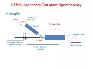
Secondary ion mass spectroscopy • It involves generating a beam of ions such as Ar+, Xe+ and Ne+ etc…., the ion beam directed into the target molecule, causing it turn to ionize.(secondary ion)
FIELDIONISATION • The method is based on behaviour of chemical compds. under high electrostatic field • When mole. is brought b/w two closely spaced electrodes in presence of high electric field(107 – 1010 v/m), it experience an electrostatic force similar to that on plates of charged condenser • If the metal surface has proper geometry (sharp tip/thin wire), the electrostatic force is sufficient to remove an electron from molecule & form ions. • Disadv – Less sensitive than electron impact method.
FIELDDESORPTION • Modified technique of field ionisation • Sample is deposited directly on anode • The electrostatic field (107 – 1010 v/m) produces ionisation as well as desorption • Useful for adsorbed species & trapped samples • Non- volatile & thermally unstable organic compds. can be easily ionised • Advantage – natural carbohydrates that do not form molecular ions by electron impact method, can be ionised by this technique
8FAST-ATOM BOMBARDMENT (FAB) • In FAB a high-energy beam of neutral atoms, typically Xe or Ar, are used Xe ions are introduce into Xegas.they producing a residual beam of energized atom (Fast Atom) • The atomic beam is produced by accelerating ions from an ion source through a charge-exchange cell. The ions pick up an electron in collisions with neutral atoms to form a beam of high energy atoms. Samples are bombarded. • FAB causes little fragmentation and usually gives a large peak corresponding to the molecular weight (molecular ion).
Benefits: rapid, simple, strong ion production.high resolution • De merits: low mol wt comp is very difficult. Multiple charged comp is dofficult.
9.Surface Ionization/Thermal ionisastion • Samples are coated with tungsten ribbon filament • Heated the filament up to evaporate the sample(2000C) • It produce the ions
10ELECTROSPRAYIONIZATION (ESI) • The ESI source consists of a very fine needle and a series of skimmers. • A sample solution is sprayed into the source chamber to form droplets. The droplets carry charge when they exit the capillary and, as the solvent evaporates (desolvation), the droplets disappear leaving highly (multiply) charged analyte molecules. • ESI is particularly useful for large biological molecules (e.g. proteins, peptides) that are difficult to vaporize or ionize, or beyond the mass range of the analyzer.
11.LASERIONIZATIONmassanalyser(LIMA) • A laser pulse vaporises a minute amount of the material from the surface of the sample, and creates a microplasma that ionizes some of the sample constituents. • The laser pulse accomplishes both vaporization and ionization of the sample. • Useful for ionisation of large biomolecules
12.ASSISTED-MATRIX LASER DESORPTION IONIZATION (AMLDI) • MALDI is an efficient desorption ionisation technique for producing gaseous ions from a solid sample by laser pulses • Macromolecules are dispersed in a solid matrix such as nicotinic acid or glycerol. • A UV laser pulse ablates the matrix which carries some of the large molecules into the gas phase in an ionized form. • MALDI is a LIMA method for vaporizing and ionizing large biological molecules (e.g., proteins, DNA fragments).
Electrostatic accelerating system • Positive ions withdrawn by electrostatic field of 400-4000 v • In accelerating chamber, the positive ions are pushed • out of the source by relatively small repeller potential, • and then accelerated by a large potential difference (1 • to 10KV - a strong electrostatic field) between the first • and second accelerating slits. • Acceleration of ions of different masses to their final • velocities.
The Magnetic Field • Accelerated particles move in curved path • Radius of curvature(r) depends on • mass of ions (m) • accelerating voltage (v) • electron charge (e) • strength of magnetic field (H)
Ion Separator / Analyser • The positively charged ions from the ion source are separated according to the respective masses of the ions under magnetic deflection. • Analyser properties • high resolution • high rate of transmission • Based on the kind of analyser, there are different types of mass spectrometers
Mass Spectrometers • Magnetic deflection mass spectrometer • Sjnglefocussing MS • Double focussing MS • Quadrupole MS • Quadrupole Mass Analyser • Quadrupole Ion-Trap Mass Analyser • Time of Flight MS • Fourior Transform Ion Cyclotron Resonance MS • (FTMS) • V. MS/MS (Tandem MS)
I. a) Single Focussing MS • Electromagnet is used to sort ions
Separation of ions is based on the principle of velocity focussing. • A small quantity of sample is injected and vaporized under high vacuum • The sample is then bombarded with electrons having 25-80 eV of energy • A valence electron is “punched” off of the molecule, and an ion is formed • Ions (+) are accelerated towards the focusing magnet
At a given potential (1 – 10 kV) each ion will have a kinetic energy: • ½ mv2 = eV • As the ions enter a magnetic field, their path is curved; the radius of the curvature is given by: • r = mv • eH • If the two equations are combined to factor out velocity: • m/e = H2r2 • 2V • Fundamental Equation for single focussing MS
At a given potential, only one mass would have the correct radius path to pass through the magnet towards the detector • “Incorrect” mass particles would strike the magnet
By varying the applied potential difference that accelerates each ion, different masses can be discerned by the focusing magnet • The detector is basically a counter, that produces a current proportional to the number of ions that strike it • This data is sent to a computer interface for graphical analysis of the mass spectrum
b) Double Focussing MS • Single focussing MS – Assumption • all ions enter the magnetic field with same KE i.e. all ions with same m/e possess same velocity • Not true in practice, ions have different initial energy & thus leave electron beam with variable energy • SFMS fails to discriminate b/w small mass differences due to small variation in KE. • To achieve better focussing, energy has to be reduced before ions are allowed to enter magnetic field
Double Focussing MS • In DFMS, electrostatic field selects & focuses ions with certain velocity / KE (slit 2) magnetic field slit 3 detector SLIT 2 BEAM OF IONS ELECTROSTATIC FIELD DETECTOR SLIT 3 MAGNETIC FIELD
Double Focussing MS • Very high resolution (90000) • However there is slow transmission of ions i.e. slow ion current. Thus this instrument is useful when high resolution is required • Determination of precise molecular weights