Download
1 / 17
300 likes | 1.97k Views
Congenital Lobar Emphysema. The case of the cyanotic Pekingese Erica Fields, DVM. Gizmo Ladd. MRN 140041 3 month old male Pekingese Acute dyspnea and cyanosis, multiple transient episodes in the last 24 hours. Radiographic Findings. Marked decrease in pulmonary vascular markings

E N D
Congenital Lobar Emphysema The case of the cyanotic Pekingese Erica Fields, DVM
Gizmo Ladd • MRN 140041 • 3 month old male Pekingese • Acute dyspnea and cyanosis, multiple transient episodes in the last 24 hours
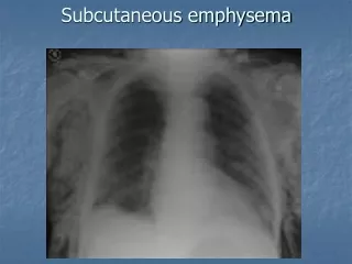
Radiographic Findings • Marked decrease in pulmonary vascular markings • Flattening of the diaphragm, consistent with hyperinflation • Thin septae throughout pulmonary parenchyma, suggestive of saccular dilations • Alveolar pattern, left caudal lung lobe—atelectasis??
Differential Diagnoses • Pulmonary hyperlucency • Tension pneumothorax • Metabolic acidosis (compensatory hyperpnea) • Pulmonary cysts (bullae) • Relative decrease in pulmonary tissue/vasculature • Air trapping • Compensatory lung expansion (as with contralateral lobectomy) • Artifact—overexposed radiograph Suter, Thoracic Radiography
Differential Diagnoses • Decreased pulmonary vascularity • Pulmonary thromboembolism (regional oligemia) • Hypovolemic shock • Myocardial disease with reduced RV output • Severe pulmonic stenosis • Addison’s disease • Pulmonary artery hypertension • Right to left shunting (reversed PDA, tetralogy of Fallot, Eisenmenger’s syndrome) Suter, Thoracic Radiography
Diagnostic Testing • Thoracic Radiographs (inspiratory and expiratory) • Lab testing, including blood gas—with CLE, often have relatively normal results • Non-selective pulmonary angiography • Nuclear medicine—Tc-MAA perfusion scanning and ventilation scanning (N-13 reported) • Computed tomography (CT) • Pulmonary function testing • Echocardiography (rule out right-to-left cardiac shunt) • Bronchoscopy (or bronchogram, as performed in one study) Amis, et al. 1987
Diagnostics—Gizmo’s case • Thoracic radiographs • CBC/Chem—mild anemia and neutrophilic leukocytosis (would expect polycythemia with RL shunt) • ECG—NSR • Blood pressure--WNL
Echocardiography—cephalic injection bubble study performed; no evidence of intracardiac shunting; however, bubbles appeared in the left atrium and ventricle after approximately 10-15 cardiac cycles—suggestive of AV malformation in pulmonary vasculature??
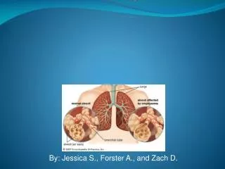
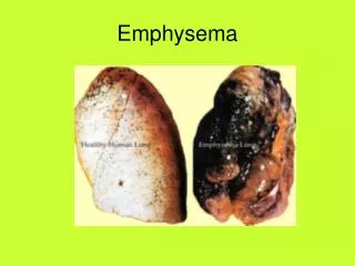
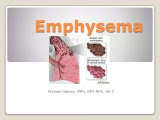
What is Congenital Lobar Emphysema? • First, what is emphysema? • Technically, it refers to destruction of alveolar walls and consequent hyperinflation of airspaces distal to the terminal bronchioli • Overinflation or air trapping can look the same, but without destruction of alveoli • Emphysema is irreversible, whereas air trapping is reversible Suter, Thoracic Radiography
Congenital Lobar Emphysema • Recognized in humans, dogs • Probably not technically emphysema in the dog, as no alveolar destruction, but the name stuck • Bronchial cartilage hypoplasia, dysplasia or aplasia • Without cartilage support, the bronchi collapse on expiration, leading to gradual air trapping and hyperinflation Amis, et al. 1987
Congenital Lobar Emphysema • In humans, boys are over-represented • Typically cranial lobes or right middle lobe • Most common presentation is dyspnea, cyanosis, and exercise intolerance • Usually recognized as infants or puppies—one case of a 5 yo Peke • Rare, but at least 3 of the reported cases are Pekingese, suggesting breed predilection Voorhout, et al. 1986
Histopathology • Usually find hypoplasia, dysplasia, or aplasia of bronchial cartilage in affected lobe • Hyperinflation of alveoli +/- alveolar destruction • Epithelium and secretory cells may be intact Billet and Sharpe. 2002
Treatment and Prognosis • Surgical excision of the affected lobe • May treat conservatively (restricted activity) if minimally affected—more experience with this in humans • In humans, good prognosis if treated surgically • In dogs, not so good—all but 1 dog in literature either died or were euthanized Billet and Sharpe, 2002. Amis, et al 1987.
References • Amis TC, Hager D, Dungworth DL, Hornof W. Congenital bronchial cartilage hypoplasia with lobar hyperinflation (congenital lobar emphysema) in an adult Pekingese. JAAHA 1987; 23: 321-329. • Billet JPHG and Sharpe A. Surgical treatment of congenital lobar emphysema in a puppy. J Sm An Pract 2002; 43: 84-87. • Suter. Thoracic Radiography. • Voorhout G, Goedegebuure SA, and Nap RC. Congenital lobar emphysema caused by aplasia of bronchial cartilage in a Pekingese puppy. Vet Pathol 1986; 23: 83-84.