Download
1 / 25
260 likes | 477 Views
Introduction to the Digestive System. For student copy. Digestive system & homeostasis. Digestive system contributes to homeostasis of body by breaking down food into forms that can be absorbed & used by body cells. also: absorbs : water, vitamins, minerals eliminates: wastes.

E N D
Introduction to the Digestive System For student copy
Digestive system & homeostasis • Digestive system contributes to homeostasis of body by breaking down food into forms that can be absorbed & used by body cells. • also: • absorbs : • water, vitamins, minerals • eliminates: • wastes
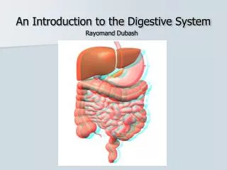
Accessory Organs 2 groups of organs: GI Tract aka Alimentary Canal continuous tube mouth anus mouth most of pharynx esophagus stomach small & large intestine teeth tongue salivary glands liver gallbladder pancreas
vocabulary • Ingestion: eating • Secretion: ~7 liters water, acids, buffers, & enzymes into lumen of GI tract • Motility: contraction/relaxation of smooth muscle in wall of GI tract mix & propel food & secretions anus • Mechanical Digestion: physically breaking down food • Chemical Digestion: • Absorption • Defecation: wastes leave body/ material defecated called feces/ gas called flatus
Layers of the gi tract (inner outer) • mucosa • submucosa • muscularis • serosa
Mucosa: 3 layers • epithelium • mouth esophagus: stratified sq. epith for protection • stomach intestines: simple columnar • cells slough off q 5-7 d • exocrine mucus glands (mucus & water) & several types endocrine glands called enteroendocrinecells interspersed
Mucosa: 2nd layer:lamina propia • areolar CT • rich in blood & lymph vessels • contains most of MALT (mucosa-associated lymphatic tissue)
Mucosa: 3rd layer:muscularis mucosa • thin layer of smooth muscle • creates small folds in epithelium increases surface area for digestion & absorption
submucosa • areolar CT that binds mucosa to muscularis • rich blood & lymphatics, glands • submucosal plexus: extensive network of neurons
Muscularis • skeletal muscle in mouth, pharynx, superior & middle parts of esophagus, external anal sphincter • voluntary swallowing & defecation • 2 sheets smooth muscle in rest of GI tract with myenteric plexus between them • outer longitudinal • inner circular
Serosa • parts of GI tract suspended in abdominopelvic cavity have this superficial layer = visceral peritoneum • esophagus lacks a serosa; has adventitia a single layer of areolar CT
Enteric Nervous System(ENS) • 100 million neurons that extends from esophagus anus • 2 plexuses: • myenteric plexus (plexus of Auerbach) • motor neurons of longitudinal & circular muscle • submucosal plexus (plexus of Meissner) • w/in submucosa supplying the secretory cells
ANS • parasympathetic fibers via X to most of GI tract (not to 2nd ½ large intestine: gets it from sacral spinal cord) • increase secretions & motility • sympathetic fibers from thoracic & upper lumbar spinal nerves • decrease secretions & motility
Peritoneum • largest serous membrane of body • simple squamous epithelium with underlying areolar CT • divided: • parietal peritoneum • visceral peritoneum
Peritoneal Cavity • space between parietal & visceral peritoneums • contains serous fluid: decreases friction • Ascites: excess serous fluid ass’c w/ some diseases
Retroperitoneal • “behind peritoneum” • kidneys & most of pancreas, end of sigmoid colon under parietal peritoneum
5 Major Peritoneal Folds • greater omentum • falciform ligament • lesser omentum • mesentery • mesocolon
Greater Omentum • largest peritoneal fold • “fatty apron” • drapes over transverse colon & coils of sm intestine • extends downward anterior to sm intestine • contains adipose cells, macrophages, plasma cells; • function to combat & contain infections
FalciformLigament • attaches liver to anterior abdominal wall & diaphragm • liver only organ in abdominopelvic cavity attached to anterior abd wall
Lesser Omentum • arises as 2 folds in serosa of stomach & duodenum
Mesentery • fold of peritoneum that attaches small intestine to posterior abdominal wall • starts @ posterior wall wraps around sm intestine reflects back to posterior abd wall • route of bld supply to sm intestine
Mesocolon • binds large intestine to posterior abd wall • route of blood supply, lymphatic drainage
Peritonitis • acute inflammation of the peritoneum: contamination by infectious microbes • from punctures of abdominal organs • trauma • surgical wounds