Download
1 / 22
441 likes | 1.13k Views
Ventilator Trouble shooting Presented by Lily To & James Lindsey. Ventilator Troubleshooting Involves identification & resolution of a technical problem A problem is a situation in which one finds oneself in that can not be immediately corrected Solving Ventilator Problems Access situation

E N D
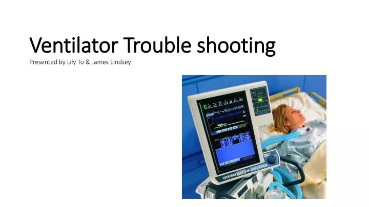
Ventilator Trouble shootingPresented by Lily To & James Lindsey
Ventilator Troubleshooting • Involves identification & resolution of a technical problem • A problem is a situation in which one finds oneself in that can not be immediately corrected • Solving Ventilator Problems • Access situation • Gather & analyze pertinent data • This information should point to a number of potential solutions • A solution should be tried – with making an observation of the patient’s response • A positive response leads to correction of the problem • A negative response – undo what was tried – find out why it didn’t work before attempting a new solution • Determining cause of the problem – helps prevent the problem from reoccurring • Protecting the Patient • Always ensure patient safety • When alarm is triggered – check patient first • Look for LOC, increased WOB, use of accessory muscles, auscultation, SpO2, heart rate, skin color, diaphoresis • Distress - bag patient, if necessary • Check alarm & alarm settings
Identifying Patient Distress • Notice when patient is “fighting the vent” or asynchrony • Signs include: tachypnea, nasal flaring , diaphoresis, use of accessory muscles, retractions, paradoxal chest abdomen movement, abnormal breath sounds, tachycardia, arrhythmia, hypertension Ventilator – Related Causes • System leaks • Disconnected circuit • Low FiO2 Patient – Ventilator asynchrony – Causes • Artificial airway problems • Bronchospasm • Secretions • Pulmonary edema • Pulmonary embolism • Dynamic hyperinflation • Abnormal respiratory drive • Body positioning • Pneumothorax • Anxiety Sudden Causes of Respiratory Distress • Improper Settings • Incorrect support mode • Sensitivity • Flow • Time cycled • PEEP
Common Patient – Related Problems • Airway problems • Kinked ET tube, biting • Displacement of tube in right lobe or upward • Rupture of an artery • Fistula obstructed ET tube • Pneumothorax • Look for increased work of breathing, nasal flaring, use of accessory muscles, • absence of breath sounds, uneven chest movement & cardiovascular assessment • Bronchospasm • Signs include: • dyspnea, wheezing, increased work of breathing, paradoxical chest/abdomen movement, • retractions and increased RAW • Secretions • Evaluation can lead to differentiate problems • Dry secretions – insufficient humidification? • Copious amounts – pulmonary edema? • Detect infection? Pneumothorax
Common Patient – Related Problems • Pulmonary Edema • Cardiogenic pulmonary edema • Sudden – thin, frothy, white to pink secretions. Follow through with additional testing – ECG, Bp, • JVD and Hx of heart disease • Treatment includes medications to reduce preload and afterload (lasix), increase contractility (Lanoxin) • Non-cardiogenic pulmonary edema • Not sudden – increase in pulmonary capillary permeability (treatment similar to above) • Dynamic Hyperinflation • Auto-PEEP causes dynamic hyperinflation – leads to difficulty triggering ventilator & increased work of breathing • Causes hypertension and reduced cardiac output • Suspected when flow does not return to baseline in flow-time curve. • Treatment: reduce TI, VE and correct RAW • Abnormalities in Respiratory Drive • Decrease is result of heavy sedation, neurological disorders, neuromuscular blockage • Increase is result of pain, anxiety, peripheral sensory stimulation, medications and improper ventilator settings
Common Patient – Related Problems • Changes in Position • Can cause accidental extubation • Alter oxygenation by bending, twisting circuit • Cause mucous plugging • Drug Induced Distress • Can cause respiratory distress & maybe failure • Abdominal Distention • Distention - can be associated with other disorders that introduce air into the stomach • (ascites, GI bleed, liver & kidney problems) • Pulmonary Embolism • Emergency • Leads to asynchrony • Sudden onset – hypoxemia • Patient presents with bilateral breath sounds, increased WOB, elevated HR, • Bp and RR • Increasing flow and FiO2 does nothing to correct • Treat with increased respiratory rate • Capnography – helps us see – reduced VT & CO2
Ventilator – Related Problems • Leaks – cuff, circuit • Alarm activates • Low/high pressure • Low minute ventilation • Inadequate Oxygenation • SpO2 alarm • Signs – hypoxemia • Inadequate Ventilator Support • Causes increased work of breathing, respiratory acidosis & hypoxemia • Leads to asynchrony • Sensitivity • Causes auto-triggering –setting too low - high pressure, patient can not trigger • Flow Setting • Air starvation – correct by increasing flow or changing flow pattern • Other Problems • Auto-PEEP – makes vent more difficult for patient to trigger a breath – correct by increasing E-time • PSV - may cause asynchrony with certain disorders and if it is set too low Puritan Bennet 840 Drager V500
Common Alarm Situations • Normal Alarm Settings: • VT: high, 200ml above setting – low, 100ml below setting • Pressure: high, 10cmH2O above PIP – low, 5cmH2O below PIP • Rate: high, 10 bpm above setting – low, 5 bpm below setting • Flow: high, 2L above setting – low, 2L below setting • Apneic: 20 seconds • FiO2: high, 5% above setting – low, 5% below setting
Common Alarm Situations Low Pressure Alarm Causes: • Patient disconnected • Circuit leaks – inspiratory/expiratory circuits • Ventilator related disconnections • Humidifiers, filters, water traps, nebulizers, closed circuit catheter • Temperature monitors • Exhalation valve leak • Cracked, unseated, improperly connected • Airway leaks • Improper cuff inflation • Cut hole in pilot balloon/ cuff • Migration of ET tube • Chest tube leaks *Most often activated by leaks* High Pressure Alarm Causes: • Coughing • Biting, kinking, positioning of ET tube • Herniation of ET tube/cuff • Increased airway resistance (secretions, edema, bronchospasm) • Decreased compliance (pneumothorax, pulmonary embolism) • Patient – ventilator asynchrony • Accumulation of water in circuit • Kinking in inspiratory circuit • Malfunction with inspiratory/expiratory valves
Additional Alarms • Low PEEP/CPAP • Activated when airway pressure falls below desired baseline during PEEP/CPAP • Causes include: leaks or by active inspiration • Apnea alarm • 20 seconds • Causes: patient apneic or disconnection, leaks, sensitivity setting • Low-Source Gas Pressure/ Power Alarm • If gas or power source fails • I:E Ratio Alarm • Most ventilators do not allow I:E ratio to be set less than 1:1 • Causes: flow set too low for desired VT delivery • I:E – may change with a change in waveform (constant to descending - lengthens TI in VC)
Additional Alarms • High PEEP/CPAP alarms • Causes are similar to those of high pressure • flow-cycle modes , check for leaks • Low VT, low VE or low flow alarms • Causes are similar to low pressure alarms • Determine if spontaneous ventilation has decreased • Check all alarms • Check flow sensors, disconnection/malfunction • High VT, high VE or high flow alarms • Check sensitivity setting, causes auto-triggering • Check patient for possible cause of increased VE • Check alarm settings • If nebulizer in use, reset alarm until treatment is completed • Check flow sensors, contamination/malfunction • Low/high FiO2 alarms • Check gas source • Check built-in oxygen analyzer is functioning properly Flow Sensor Nebulizer
Flow-Volume • Use of Ventilator Graphics to Identify Ventilator Problems • Ventilator graphics can alert of abnormalities before • obvious signs appear • Flow-time & Pressure-time graphs are used for accessing • patient triggering, flow starvation, auto-PEEP, I:E time, • flow pattern, plateau time, rise times and asynchrony • Volume-time graph accesses auto-PEEP • Pressure-Volume loop accesses leaks, overdistention, • increased RAW, asynchrony and patient triggering • Flow-Volume loops are used to access • obstructive/restrictive lungs, the effects of bronchodilators • and leaks • Waveform ringing in Flow-time & Pressure-time • Occurs when flow & pressure are very high at a beginning • of a breath – a result of oscillation of air at beginning of a • breath
Use of Ventilator Graphics to ID ProblemsLeaks – low pressure, low volume , low minute ventilation or apnea will trigger alarm Pressure-Volume Loop Flow-Volume Loop Leak Leak Flow-time curve Volume-time curve Auto-PEEP, air trapping
Examples of additional graphic curves Pressure-Volume Loop Obstruction: administer bronchodilator Overdistention Overdistention Correct: increase E-time Correct: reduce volume, pressure
Unexpected Ventilator Responses • Unseated/Obstructed Expiratory Valve • Blocked or unseated valve, unable to get expiratory pause – plateau pressure • High Tidal Volume Delivery • Occurs with small volume nebulizer (SVN) • Flowmeters can add extra flow – can increase tidal volume • Excessive CPAP/PEEP • Eliminate leaks – causes application of high flow to maintain CPAP/PEEP • Nebulizer Impairment of Patient’s Ability to Trigger PSV • Nebulizer makes it more difficult for patient to trigger ventilator • Usually occurs with external gas sourced nebulizer • Use manufacturer’s nebulizer if provided Flowmeter 840
Increased VT, VE or rate alarm Please Notealways start by checking patient’s stability and is adequately ventilated N o Is patient demand VE increased Check cause of increased VE demand to determine if change is needed yes yes Is vent Auto-triggering 1. Check sensitivity setting 2. Check the MMV setting yes Is a nebulizer In use Adjust vent settings until treatment is completed Is flow sensor malfunctioning yes Clean & calibrate sensor Clear sensor line Check its function and replace if needed yes Is alarm set too low Adjust alarm setting 1. Check machine for sensitivity level for auto-triggering 2. Check for cause of increased VE 3. Ensure alarms have been properly set 4. External nebulizer used; reset alarm until treatment is completed 5. Check flow sensors for calibrations, contamination or malfunction Check operators Manual/contact manufacturer
Low pressure. Low PEEP, low VT, low VE Is patient disconnected yes Reconnect N o yes Is there a leak in the circuit Repair/replace circuit yes Reinflate cuff/check it’s pressure –replace tube if necessary Is there a cuff leak yes Is there a chest tube leak Contact physician/monitor pt yes Clear the line Is proximal airway pressure line obstructed Clear sensor & recalibrate it Clear sensor line & recheck Check sensor function & replace sensor if necessary Is the flow sensor malfunctioning yes Alarm set inappropriately yes Reset 1. Check for disconnection 2. Check for leaks in ventilator, circuits, airway & chest tubes 3. Check proximal pressure line is connected & unobstructed 4. Low-pressure maybe accompanied by a low minute volume or low tidal volume alarm Check manual/contact trained specialist
High pressure, High PEEP alarms No Is artificial airway completely obstructed Can it be cleared Change artificial airway yes Suction or relieve irritation Is pt coughing yes Suction pt Are there secretions in the airway yes Drain condensation 3. kinks in ventilator circuit Check water traps Is the circuit obstructed yes Is ET tube being bitten yes Insert a bite block Is the position of artificial airway altered yes Reposition artificial airway Is the Raw increased or compliance increased Assess & Correct Secretions 5. Pulmonary edema Bronchospasm 6. Pneumothorax Mucosal edema 7. Pleural effusion Pneumonia 8. Other yes Continued
Continued - High pressure, High PEEP Is pt breathing asynchronously Check inspiratory gas flow 4. Check mode of ventilation Check sensitivity 5. Consider sedation Check vent parameters yes No yes Check & treat for increased Raw (suction, bronchodilator) Increase flow to shorten Ti and increase TE Decrease VE Auto-PEEP present Is exhalation valve malfunctioning Fix or replace valve yes Reduce pressure yes Is the venting pressure too high Increase alarm setting Is alarm set too low yes 1. Pt coughing; determine if secretions have built up in airway or pt is biting ET tube 2. Check for kinks or displacement of ET tube and circuit 3. Check to see if RAW has increased or CL has decreased 4. Check is patient is breathing synchronously with vent 5. Determine if there is auto-PEEP has developed 6. Make sure the expiratory filter & expiratory valve are functioning properly. Check for possible causes ET cuff blocking the end of the artificial airway
I:E Indicator Is an adverse ratio desired yes Activate inverse ratio No Decrease inspiratory time yes Is vent time cycled yes Is volume being used with set flow too low Increase flow yes Decrease volume Is volume being used with a set volume too high yes Is the rate too high Decrease rate Is vent flow reduced due to mechanical problem, increased Raw, or decreased compliance yes Eval patient & vent’s performance and correct problem 1. Usually indicates I:E ratio greater than 1:1 2. If inverse is goal: disable I:E ratio limit or ignore alarm 3. If normal I:E desired: check alarm If increased RAW/decreased CL has resulted in lower flow, tx cause If flow is too low for desired VT, increase flow or change waveform Change mode or VE parameters
Apneic Alarm No Is an actual apneic episode occurring Readjust vent support yes Is the alarm setting appropriate yes Reset alarm yes Reset the sensitivity Is vent insensitive to patient effort Is there a leak See low pressure alarms yes Is flow or pressure sensor faulty Clean recalibrate, check & replace sensor if necessary yes Check operator’s manual/contact trained technician 1. Is patient apneic 2. Check for leaks 3. Check sensitivity to make sure vent can detect patient effort 4. Check alarm time interval and volume setting
References: • AARC Clinical Practice Guidelines • Basic Clinical Lab Competencies for Respiratory Care, 5th Ed., White • Cardiopulmonary Anatomy & Physiology, Essentials of Respiratory Care, 6th Ed, Des Jardins • Egan’s Fundamentals of Respiratory Care, 10th Ed, Kacmarek, Stoller, Heuer • emedicine.com • Equipment Theory for Respiratory Care, 4th Ed., White • John Hopkins Medical Health Library, hopkinsmedicine.org • MayoClinic.com • Mechanical Ventilation Physiological and Clinical Applications, 5th Ed 2014, Pilbeam • Medline Plus, 2013 • Medscape • NCBI, National Center for Biotechnology Information, U.S. National Library of Medicine, 2013 • NDNR, Naturopathic Doctor News & Review, 2013 • RC Journal • Respiratory Care, Principles & Practice, 2nd Ed, Hess • The Essentials of Respiratory Care, 4th Ed, Kacmarek