Download
1 / 1
10 likes | 105 Views
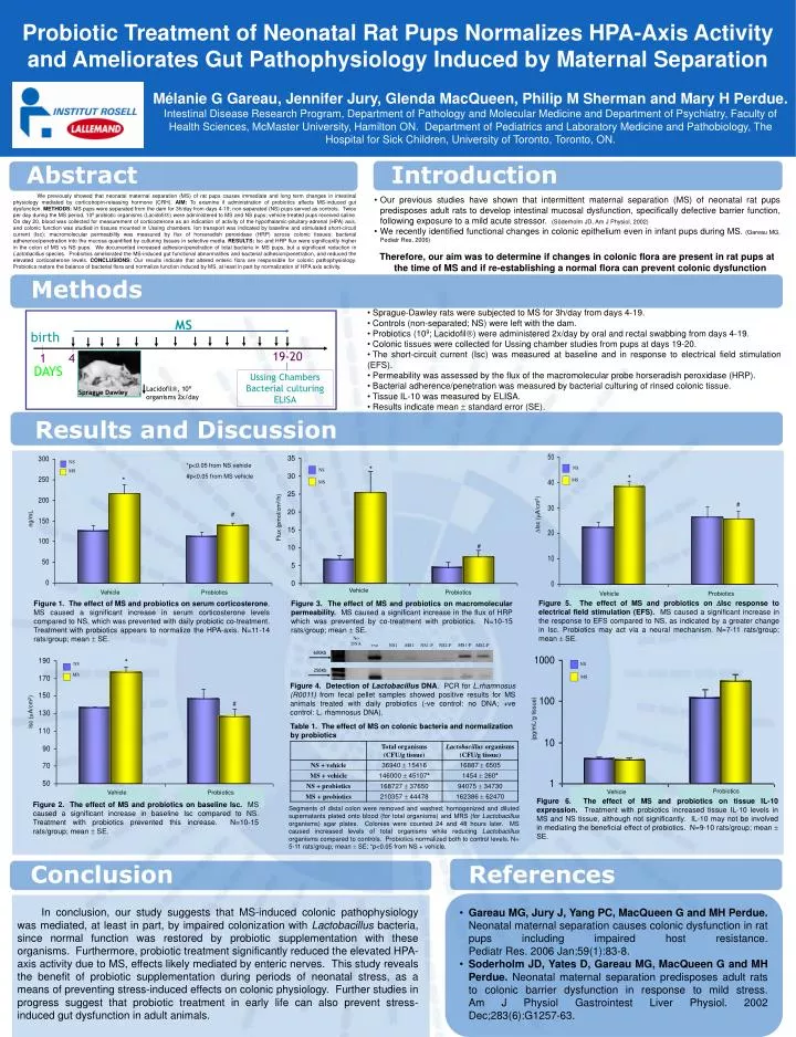
No DNA. MS1-P. +ve. NS1. MS1. NS1-P. NS2-P. MS2-P. 600Kb. 250Kb. MS. birth. 19-20. 1. 4. DAYS. Ussing Chambers Bacterial culturing ELISA. Lacidofil , 10 9 organisms 2x/day. Sprague Dawley. *. NS. MS. Flux ( pmol /cm 2 /h). NS. *. MS. #. #. Isc ( A/cm 2 ). NS.

E N D
No DNA MS1-P +ve NS1 MS1 NS1-P NS2-P MS2-P 600Kb 250Kb MS birth 19-20 1 4 DAYS Ussing Chambers Bacterial culturing ELISA Lacidofil, 109 organisms 2x/day Sprague Dawley * NS MS Flux (pmol/cm2/h) NS * MS # # Isc (A/cm2) NS MS * Vehicle Probiotics # ng/mL Vehicle Probiotics Vehicle Probiotics NS MS * NS MS (pg/mL/g tissue) # Isc (A/cm2) Probiotics Vehicle Vehicle Probiotics Probiotic Treatment of Neonatal Rat Pups Normalizes HPA-Axis Activity and Ameliorates Gut Pathophysiology Induced by Maternal Separation Mélanie G Gareau, Jennifer Jury, Glenda MacQueen, Philip M Sherman and Mary H Perdue. Intestinal Disease Research Program, Department of Pathology and Molecular Medicine and Department of Psychiatry, Faculty of Health Sciences, McMaster University, Hamilton ON. Department of Pediatrics and Laboratory Medicine and Pathobiology, The Hospital for Sick Children, University of Toronto, Toronto, ON. Abstract Introduction We previously showed that neonatal maternal separation (MS) of rat pups causes immediate and long term changes in intestinal physiology mediated by corticotropin-releasing hormone (CRH). AIM: To examine if administration of probiotics affects MS-induced gut dysfunction. METHODS: MS pups were separated from the dam for 3h/day from days 4-19; non-separated (NS) pups served as controls. Twice per day during the MS period, 109 probiotic organisms (Lacidofil) were administered to MS and NS pups; vehicle-treated pups received saline. On day 20, blood was collected for measurement of corticosterone as an indication of activity of the hypothalamic-pituitary-adrenal (HPA) axis, and colonic function was studied in tissues mounted in Ussing chambers. Ion transport was indicated by baseline and stimulated short-circuit current (Isc); macromolecular permeability was measured by flux of horseradish peroxidase (HRP) across colonic tissues; bacterial adherence/penetration into the mucosa quantified by culturing tissues in selective media. RESULTS: Isc and HRP flux were significantly higher in the colon of MS vs NS pups. We documented increased adhesion/penetration of total bacteria in MS pups, but a significant reduction in Lactobacillus species. Probiotics ameliorated the MS-induced gut functional abnormalities and bacterial adhesion/penetration, and reduced the elevated corticosterone levels. CONCLUSIONS: Our results indicate that altered enteric flora are responsible for colonic pathophysiology. Probiotics restore the balance of bacterial flora and normalize function induced by MS, at least in part by normalization of HPA axis activity. • Our previous studies have shown that intermittent maternal separation (MS) of neonatal rat pups predisposes adult rats to develop intestinal mucosal dysfunction, specifically defective barrier function, following exposure to a mild acute stressor.(Söderholm JD, Am J Physiol, 2002) • We recently identified functional changes in colonic epithelium even in infant pups during MS. (Gareau MG, Pediatr Res, 2006) • Therefore, our aim was to determine if changes in colonic flora are present in rat pups at the time of MS and if re-establishing a normal flora can prevent colonic dysfunction Methods • Sprague-Dawley rats were subjected to MS for 3h/day from days 4-19. • Controls (non-separated; NS) were left with the dam. • Probiotics (109; Lacidofil) were administered 2x/day by oral and rectal swabbing from days 4-19. • Colonic tissues were collected for Ussing chamber studies from pups at days 19-20. • The short-circuit current (Isc) was measured at baseline and in response to electrical field stimulation (EFS). • Permeability was assessed by the flux of the macromolecular probe horseradish peroxidase (HRP). • Bacterial adherence/penetration was measured by bacterial culturing of rinsed colonic tissue. • Tissue IL-10 was measured by ELISA. • Results indicate mean standard error (SE). Results and Discussion *p<0.05 from NS vehicle #p<0.05 from MS vehicle Figure 5.The effect of MS and probiotics on Isc response to electrical field stimulation (EFS). MS caused a significant increase in the response to EFS compared to NS, as indicated by a greater change in Isc. Probiotics may act via a neural mechanism. N=7-11 rats/group; mean SE. Figure 3.The effect of MS and probiotics on macromolecular permeability. MS caused a significant increase in the flux of HRP which was prevented by co-treatment with probiotics. N=10-15 rats/group; mean SE. Figure 1.The effect of MS and probiotics on serum corticosterone. MS caused a significant increase in serum corticosterone levels compared to NS, which was prevented with daily probiotic co-treatment. Treatment with probiotics appears to normalize the HPA-axis. N=11-14 rats/group; mean SE. Figure 4.Detection of Lactobacillus DNA. PCR for L.rhamnosus (R0011) from fecal pellet samples showed positive results for MS animals treated with daily probiotics (-ve control: no DNA; +ve control: L. rhamnosus DNA). Table 1. The effect of MS on colonic bacteria and normalization by probiotics Figure 6.The effect of MS and probiotics on tissue IL-10 expression. Treatment with probiotics increased tissue IL-10 levels in MS and NS tissue, although not significantly. IL-10 may not be involved in mediating the beneficial effect of probiotics. N=9-10 rats/group; mean SE. Figure 2.The effect of MS and probiotics on baseline Isc. MS caused a significant increase in baseline Isc compared to NS. Treatment with probiotics prevented this increase. N=10-15 rats/group; mean SE. Segments of distal colon were removed and washed; homogenized and diluted supernatants plated onto blood (for total organisms) and MRS (for Lactobacillus organisms) agar plates. Colonies were counted 24 and 48 hours later. MS caused increased levels of total organisms while reducing Lactobacillus organisms compared to controls. Probiotics normalized both to control levels. N= 5-11 rats/group; mean ± SE; *p<0.05 from NS + vehicle. Conclusion References In conclusion, our study suggests that MS-induced colonic pathophysiology was mediated, at least in part, by impaired colonization with Lactobacillus bacteria, since normal function was restored by probiotic supplementation with these organisms. Furthermore, probiotic treatment significantly reduced the elevated HPA-axis activity due to MS, effects likely mediated by enteric nerves. This study reveals the benefit of probiotic supplementation during periods of neonatal stress, as a means of preventing stress-induced effects on colonic physiology. Further studies in progress suggest that probiotic treatment in early life can also prevent stress-induced gut dysfunction in adult animals. • Gareau MG, Jury J, Yang PC, MacQueen G and MH Perdue. Neonatal maternal separation causes colonic dysfunction in rat pups including impaired host resistance.Pediatr Res. 2006 Jan;59(1):83-8. • Soderholm JD, Yates D, Gareau MG, MacQueen G and MH Perdue. Neonatal maternal separation predisposes adult rats to colonic barrier dysfunction in response to mild stress.Am J Physiol Gastrointest Liver Physiol. 2002 Dec;283(6):G1257-63.