Download
1 / 78
780 likes | 917 Views
THE NERVOUS SYSTEM PART 2. CHAPTER 11. Neurophysiology. Neurons are highly irritable Action potentials, or nerve impulses, are: Electrical impulses carried along the length of axons Always the same regardless of stimulus The underlying functional feature of the nervous system.

E N D
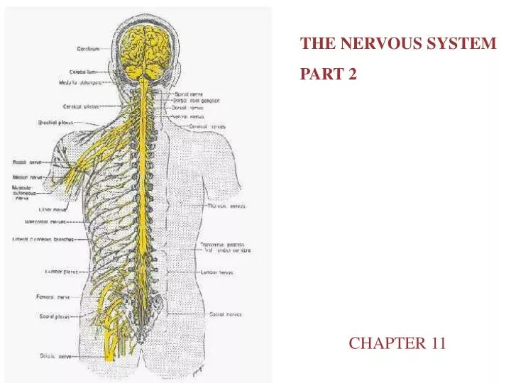
THE NERVOUS SYSTEM PART 2 CHAPTER 11
Neurophysiology • Neurons are highly irritable • Action potentials, or nerve impulses, are: • Electrical impulses carried along the length of axons • Always the same regardless of stimulus • The underlying functional feature of the nervous system
Electricity Definitions • Voltage (V) – measure of potential energy generated by separated charge • Potential difference – voltage measured between two points • Current (I) – the flow of electrical charge between two points • Resistance (R) – hindrance to charge flow • Insulator – substance with high electrical resistance • Conductor – substance with low electrical resistance
Electrical Current and the Body • Reflects the flow of ions rather than electrons • Na+ K+ • There is a potential on either side of membranes when: • The number of ions is different across the membrane • The membrane provides a resistance to ion flow
Role of Ion Channels • Types of plasma membrane ion channels: • Passive, or leakage, channels – always open • Chemically gated channels – open with binding of a specific neurotransmitter • Voltage-gated channels – open and close in response to membrane potential • Mechanically gated channels – open and close in response to physical deformation of receptors
Operation of a Gated Channel • Example: Na+-K+ gated channel • Closed when a neurotransmitter is not bound to the extracellular receptor • Na+ cannot enter the cell and K+ cannot exit the cell • Open when a neurotransmitter is attached to the receptor • Na+ enters the cell and K+ exits the cell
Operation of a Gated Channel Figure 11.6a
Operation of a Voltage-Gated Channel • Example: Na+ channel • Closed when the intracellular environment is negative • Na+ cannot enter the cell • Open when the intracellular environment is positive • Na+ can enter the cell
Operation of a Voltage-Gated Channel Figure 11.6b
Gated Channels • When gated channels are open: • Ions move quickly across the membrane • Movement is along their electrochemical gradients • An electrical current is created • Voltage changes across the membrane
Electrochemical Gradient • Ions flow along their chemical gradient when they move from an area of high concentration to an area of low concentration • Ions flow along their electrical gradient when they move toward an area of opposite charge • Electrochemical gradient – the electrical and chemical gradients taken together
Resting Membrane Potential (Vr) • The potential difference (–70 mV) across the membrane of a resting neuron • It is generated by different concentrations of Na+, K+, Cl, and protein anions (A) • Ionic differences are the consequence of: • Differential permeability of the neurilemma to Na+ and K+ • Operation of the sodium-potassium pump
Resting Membrane Potential (Vr) The resulting net outward diffusion of positive charged K+ leads to a state of relative negativity on the inner membrane face. Sodium-potassium pump transports 3 Na+ out for each K+ back into the cell. Figure 11.8
Membrane Potentials: Signals • Used to integrate, send, and receive information • Membrane potential changes are produced by: • Changes in membrane permeability to ions • Alterations of ion concentrations across the membrane • Types of signals – graded potentials and action potentials
Changes in Membrane Potential • Changes are caused by three events • Depolarization – the inside of the membrane becomes less negative • Repolarization – the membrane returns to its resting membrane potential • Hyperpolarization – the inside of the membrane becomes more negative than the resting potential
Changes in Membrane Potential Figure 11.9
Graded Potentials • Short-lived, local changes in membrane potential • Decrease in intensity with distance • Their magnitude varies directly with the strength of the stimulus • Sufficiently strong graded potentials can initiate action potentials
Graded Potentials Figure 11.10
Graded Potentials • Voltage changes in graded potentials are decremental • Current is quickly dissipated due to the leaky plasma membrane • Can only travel over short distances
Graded Potentials Figure 11.11
Action Potentials (APs) • A brief reversal of membrane potential with a total amplitude of 100 mV • Action potentials are only generated by muscle cells and neurons • They do not decrease in strength over distance • They are the principal means of neural communication • An action potential in the axon of a neuron is a nerve impulse
Action Potential: Resting State • Na+ and K+ channels are closed • Leakage accounts for small movements of Na+ and K+ • Each Na+ channel has two voltage-regulated gates • Activation gates – closed in the resting state • Inactivation gates – open in the resting state Figure 11.12.1
Action Potential: Depolarization Phase • Na+ permeability increases; membrane potential reverses • Na+ gates are opened; K+ gates are closed • Threshold – a critical level of depolarization (-55 to -50 mV) • At threshold, depolarization becomes self-generating Figure 11.12.2
Action Potential: Repolarization Phase • Sodium inactivation gates close • Membrane permeability to Na+ declines to resting levels • As sodium gates close, voltage-sensitive K+ gates open • K+ exits the cell and internal negativity of the resting neuron is restored Figure 11.12.3
Action Potential: Hyperpolarization • Potassium gates remain open, causing an excessive efflux of K+ • This efflux causes hyperpolarization of the membrane (undershoot) • The neuron is insensitive to stimulus and depolarization during this time Figure 11.12.4
Action Potential: Role of the Sodium-Potassium Pump • Repolarization • Restores the resting electrical conditions of the neuron • Does not restore the resting ionic conditions • Ionic redistribution back to resting conditions is restored by the sodium-potassium pump
Phases of the Action Potential • 1 – resting state • 2 – depolarization phase • 3 – repolarization phase • 4 – hyperpolarization
Threshold and Action Potentials • Threshold – membrane is depolarized by 15 to 20 mV • Established by the total amount of current flowing through the membrane • Weak (subthreshold) stimuli are not relayed into action potentials • Strong (threshold) stimuli are relayed into action potentials • All-or-none phenomenon – action potentials either happen completely, or not at all
Absolute Refractory Period • Time from the opening of the Na+ activation gates until the closing of inactivation gates • The absolute refractory period: • Prevents the neuron from generating an action potential • Ensures that each action potential is separate • Enforces one-way transmission of nerve impulses
Absolute Refractory Period Figure 11.15
Relative Refractory Period • The interval following the absolute refractory period when: • Sodium gates are closed • Potassium gates are open • Repolarization is occurring • The threshold level is elevated, allowing strong stimuli to increase the frequency of action potential events
Nerve Fiber Classification • Nerve fibers are classified according to: • Diameter • Degree of myelination • Speed of conduction
Synapses • A junction that mediates information transfer from one neuron: • To another neuron • To an effector cell • Presynaptic neuron – conducts impulses toward the synapse • Postsynaptic neuron – transmits impulses away from the synapse
Types of Synapses • Axodendritic – synapses between the axon of one neuron and the dendrite of another • Axosomatic – synapses between the axon of one neuron and the soma of another • Other types of synapses include: • Axoaxonic (axon to axon) • Dendrodendritic (dendrite to dendrite) • Dendrosomatic (dendrites to soma)
Electrical Synapses • Electrical synapses: • Are less common than chemical synapses • Correspond to gap junctions found in other cell types • Are important in the CNS in: • Arousal from sleep • Mental attention • Emotions and memory • Ion and water homeostasis
Chemical Synapses • Specialized for the release and reception of neurotransmitters • Typically composed of two parts: • Axonal terminal of the presynaptic neuron, which contains synaptic vesicles • Receptor region on the dendrite(s) or soma of the postsynaptic neuron
Synaptic Cleft • Fluid-filled space separating the presynaptic and postsynaptic neurons • Prevents nerve impulses from directly passing from one neuron to the next • Transmission across the synaptic cleft: • Is a chemical event (as opposed to an electrical one) • Ensures unidirectional communication between neurons
Synaptic Cleft: Information Transfer • Nerve impulses reach the axonal terminal of the presynaptic neuron and open Ca2+ channels • Neurotransmitter is released into the synaptic cleft via exocytosis in response to synaptotagmin • Neurotransmitter crosses the synaptic cleft and binds to receptors on the postsynaptic neuron • Postsynaptic membrane permeability changes, causing an excitatory or inhibitory effect
Termination of Neurotransmitter Effects • Neurotransmitter bound to a postsynaptic neuron: • Produces a continuous postsynaptic effect • Blocks reception of additional “messages” • Must be removed from its receptor • Removal of neurotransmitters occurs when they: • Are degraded by enzymes • Are reabsorbed by astrocytes or the presynaptic terminals • Diffuse from the synaptic cleft
Synaptic Delay • Neurotransmitter must be released, diffuse across the synapse, and bind to receptors • Synaptic delay – time needed to do this (0.3-5.0 ms) • Synaptic delay is the rate-limiting step of neural transmission
Neurotransmitters • Chemicals used for neuronal communication with the body and the brain • 50 different neurotransmitters have been identified • Classified chemically and functionally
Chemical Neurotransmitters • Acetylcholine (ACh) • Biogenic amines • Amino acids • Peptides • Novel messengers: ATP and dissolved gases NO and CO
Neurotransmitters: Acetylcholine • First neurotransmitter identified, and best understood • Released at the neuromuscular junction • Synthesized and enclosed in synaptic vesicles • Degraded by the enzyme acetylcholinesterase (AChE) • Released by: • All neurons that stimulate skeletal muscle • Some neurons in the autonomic nervous system
Functional Classification of Neurotransmitters • Two classifications: excitatory and inhibitory • Excitatory neurotransmitters cause depolarizations (e.g., glutamate) • Inhibitory neurotransmitters cause hyperpolarizations (e.g., GABA and glycine)
Functional Classification of Neurotransmitters • Some neurotransmitters have both excitatory and inhibitory effects • Determined by the receptor type of the postsynaptic neuron • Example: acetylcholine • Excitatory at neuromuscular junctions with skeletal muscle • Inhibitory in cardiac muscle