Download
1 / 57
570 likes | 577 Views
Chapter 9 The Autonomic Nervous System. Autonomic Motor Nerves. Page 244. Motor (efferent) division: Two subdivisions 1.Somatic nervous system 軀體神經系統 = voluntary 2.Autonomic nervous system 自主神經系統 = involuntary. Somatic vs. Autonomic System. Page 245.

E N D
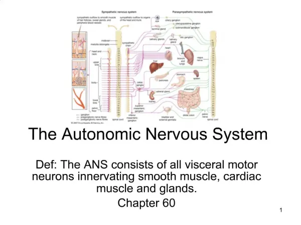
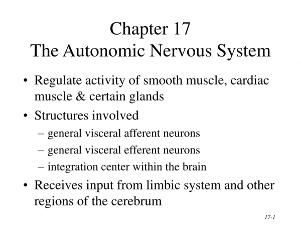
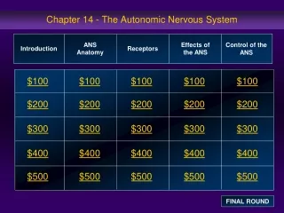
Chapter 9 The Autonomic Nervous System
Autonomic Motor Nerves Page 244 • Motor (efferent) division: Two subdivisions 1.Somatic nervous system 軀體神經系統 = voluntary 2.Autonomic nervous system 自主神經系統 = involuntary
Somatic vs. Autonomic System Page 245
Somatic vs. Autonomic System Page 244
Autonomic Neurons Page 244 • Somatic motor neurons have cell bodies in the spinal cord and just one neuron traveling from spinal cord to effector. • The autonomic motor system has two sets of neurons in the PNS. • The first has cell bodies in the brain or spinal cord and synapses in an autonomic ganglion. • The second has cell bodies in the ganglion and synapses on the effector.
Autonomic Ganglia • Located in the head, neck, and abdomen as well as in chains along either side of the spinal cord Page 244
Visceral Effector Organs Page 245 • Somewhat independent of innervation and will not atrophy if a nerve is cut (unlike skeletal muscle) • Cardiac muscle and some smooth muscle contract rhythmically without nerve stimulation. Autonomic innervation can speed up or slow down intrinsic contractions. • Unlike somatic motor neurons (which are always stimulatory), autonomic motor neurons can stimulate or inhibit.
Autonomic Neurons Page 246 Preganglionic neurons: originate in the midbrain or hindbrain or from the thoracic, lumbar, or sacral spinal cord Postganglionic neurons: originate in ganglion
Page 245 Comparison of Somatic and Autonomic Nervous Systems
Neurotransmitters • Somatic motor neurons release only acetylcholine which is always excitatory. • Autonomic neurons release mainly acetylcholine and norepinephrine but may be excitatory or inhibitory Page 245
Parasympathetic vs Sympathetic Neurons Parasympathetic Division Page 246 Short Postganglionic axon Ganglionic neuron Preganglionic neuron Long preganglionic axon Autonomic ganglion Sympathetic Division Short, branching preganglionic axon Long postganglionic axon Preganglionic neuron Ganglionic neuron Autonomic ganglion
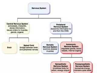
Page 249 SympatheticandParasympatheticDivisions; the solid lines indicate preganglionic fibers, and the dashed lines indicate postganglionic fibers
Sympathetic Divisions Eye Blood vessels and Sweat glands of head Page 249 Salivary glands Blood vessels Heart Cardiac and pulmonary plexuses Superior cervical ganglion Middle cervical ganglion Inferior cervical ganglion T1 T1 T1 Lung Celiac ganglion Greater thoracic splanchnic nerve T2 T2 Liver and gallbladder Lesser thoracic splanchnic nerve Stomach T3 T3 Spleen T4 T4 Adrenal medulla Superior mesenteric ganglion T5 T5 Kidney T6 T6 Pancreas T7 T7 Large intestine T8 T8 Small intestine T9 T9 T10 T10 Inferior mesenteric ganglion T11 T11 Postganglionic fibers to skin, blood vessels Rectum T12 T12 L1 L1 L2 L2 L2 Least thoracic splanchnic nerve L3 Bladder Hypogastric plexus Lumbar splanchnic nerves L4 Spinal cord L5 Vas deferens Sacral splanchnic nerves Seminal vesicle S1 Prostate Sympathetic chain ganglia S2 Ovary Uterus
Sympathetic Division Page 246 • Preganglionic neurons come from the thoracic and lumbar regions of the spinal cord. • Also called the thoracolumbar division • They synapse in sympathetic ganglia that run parallel to the spinal cord. • These are called the paravertebral ganglia. • These ganglia are connected, forming a sympathetic chain of ganglia.
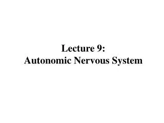
Ciliary ganglion Lacrimal gland Pterygopalatine ganglion Parotid salivary gland CN III Submandibular salivary gland Sublingual salivary gland CN VII Submanibular ganglion Page 249 CN IX Otic ganglion Pons Heart CN X Cardiac plexus Pulmonary plexus Esophageal plexus Lung Esophagus Liver Abdominal Aortic plexus Gallbladder Stomach Spleen Kidney Spinal cord Ureter Pancreas Small intestine Hypogastric plexus Descending colon Rectum S2 S3 S4 Pelvic splanchnic nerves Bladder Penis Scrotum Ovary Uterus
Autonomic Nervous System Page 248 & 249
Sympathetic Preganglionic Neurons Page 246
Sympathetic Neuron Pathways Page 247
Types of Sympathetic Pathways Preganglionic axon Postganglionic axon Posterior root ganglion Blood vessel Posterior root Hair White ramus Anterior root Lateral horn Cardiac plexus (parasympathetic fibers of plexus not shown) Arrector pili and sweat glands Spinal nerve Gray ramus White ramus Heart Sympathetic trunk ganglion Sympathetic trunk (b) Postganglionic sympathetic nerve pathway (a) Spinal nerve pathway
Types of Sympathetic Pathways Sympathetic trunk ganglion Gray ramus White ramus communicans White ramus Splanchnic nerve Adrenal medulla Preganglionic axon Splanchnic nerves Prevertebral ganglion Prevertebral ganglion (no synapse occurs) Intestine (c) Splanchnic nerve pathway (d) Adrenal medulla pathway
Collateral (Prevertebral) Sympathetic Ganglia Page 248 *The prevertebral ganglia differ from the sympathetic trunk ganglia.
Greater thoracic splanchnic nerve Trachea Left vagus nerve (X) Sympathetic trunk ganglion Right vagus nerve (X) Cardiac plexus Greater thoracic splanchnic nerve Pulmonary plexus Esophageal plexus Lesser thoracic splanchnic nerve Aorta Esophagus Diaphragm Celiac ganglia and plexus Celiac trunk (artery) Superior mesenteric artery Superior mesenteric ganglia and plexus Abdominal aortic plexus Inferior mesenteric ganglia and plexus Inferior mesenteric artery Hypogastric plexus
Collateral Ganglia Page 247 • Many of the sympathetic neurons that exit the spinal cord below the diaphragm do not synapse in the sympathetic chain of ganglia. • Instead, they form splanchnic nerves, which synapse in collateral ganglia. • Collateral ganglia include celiac, superior mesenteric, and inferior mesenteric ganglia. • Postganglionic neurons innervate organs of the digestive, urinary, and reproductive systems.
Adrenal Glands Page 247 • The adrenal medulla secretes epinephrine and norepinephrine when stimulated by the sympathetic nervous system. • Embryologically, the adrenal medulla is a modified ganglion and is innervated directly by preganglionic sympathetic neurons.
Parasympathetic Division • Preganglionic neurons come from the brain or sacral region of the spinal cord. • Also called the craniosacral division • They synapse on ganglia located near or in effector organs. • Called terminal ganglia Page 247 & 248
Autonomic Functioning Page 247 • Sympathetic – “fight-or-flight” • Response to unusual stimulus • Takes over to increase activities • Remember as the “E” division = exercise, excitement, emergency, and embarrassment
Autonomic Functioning Page 247 • Parasympathetic – housekeeping activites • Conserves energy • Maintains daily necessary body functions • Remember as the “D” division - digestion, defecation, and diuresis
Page 251 Neurotransmitters
Adrenergic Synaptic Transmission Page 253
α and β Adrenergic Receptors • Two types of α (α1 and α2) • Two types of β (β1 and β2) • All act using G-proteins and second messenger systems. • β receptors use cAMP. • α receptors use a Ca2+ second messenger system. Page 253
α and β Adrenergic Receptors • Adrenergic effects in different organs Page 254
Response to Adrenergic Stimulation Page 252 • Can stimulate or inhibit, depending on receptors • Stimulation: heart, dilatory muscles of the iris, smooth muscles of some blood vessels (causes vessel constriction) • Inhibition: Bronchioles in lungs, other blood vessels; inhibits contraction and causes dilation of these structures
Response to Cholinergic Stimulation • ACh released from preganglionic neurons of both the sympathetic and parasympathetic division is stimulatory. • ACh from postganglionic neurons of the parasympathetic division can be stimulatory or inhibitory, depending on receptors. Page 256
Two Classes of Acetylcholine Receptors Page 257 • Nicotinic: found in autonomic ganglia • Stimulated by ACh • Serve as ion channels • 270kD • consists of a, b, g and d • Each a-subunit possesses a binding site for acetylcholine
Nicotinic Receptor Page 257
Two Classes of Acetylcholine Receptors • Muscarinic: found in visceral organs • Five types identified; can be stimulatory or inhibitory (opening K+ or Ca2+ channels) • Use G-proteins and second messenger system • 70kD glycoprotein • 7TMS family of receptor Page 257
Muscarinic Receptor Page 257
ACh Receptor Structure Page 257
ACh Receptor Function Page 257
Other Autonomic Neurotransmitters • Some postganglionic autonomic neurons are not inactivated by drugs that block ACh or norepinephrine activity. • Called “nonadrenergic, noncholinergic fibers” • Proposed neurotransmitters include ATP, vasoactive intestinal peptide, and nitric oxide. Page 258
Page 258 (sildenafil) Viagra 威而鋼 phosphodiesterase GMP *NO can also produce smooth muscle relaxation in the stomach, intestines, and urinary bladder.
Organs with Dual Innervation • Most visceral organs are innervated by both sympathetic and parasympathetic neurons. • Most of the time these systems are antagonists: • Heart rate • Digestive functions • Pupil diameter Page 258
Cooperative Effects Page 258 • Occur when both divisions produce different effects that work together to promote a single action: • Erection and ejaculation: Parasympathetic division causes vasodilation and erection; sympathetic causes ejaculation • Urination: Parasympathetic division aids in urinary bladder contraction; sympathetic helps with bladder muscle tone to control urination.
Complementary Effects • Occur when both divisions produce similar effects on the same target • Salivary gland secretion: Parasympathetic division stimulates secretion of watery saliva; sympathetic constricts blood vessels so the secretion is thicker. Page 258