Download
1 / 117
1.17k likes | 1.33k Views
NASOPHARYNX, PNS & SALIVARY GLANDS. DR. SRINIVAS RAJKUMAR THIRAVIARAJ. Why is this subdivision necessary?. The primary tumors in each of these areas have different routes of • spread • nodal dissemination • prognosis. Cuboidal chamber Begins at Posterior Choana

E N D
NASOPHARYNX, PNS & SALIVARY GLANDS DR. SRINIVAS RAJKUMAR THIRAVIARAJ
Why is this subdivisionnecessary? The primary tumors in each of these areas have different routes of • spread • nodal dissemination • prognosis
Cuboidal chamber • Begins at Posterior Choana • Continues into Oropharynx via pharyngeal isthmus
Boundaries: Anterior: • posterior nasal cavity Posterosuperior: • Lower clivus, upper cervical • spine, and prevertebral • muscles Inferior: • Divided from the oropharynx • by a horizontal line drawn • along the hard and soft • palates
Level I • below mylohyoid muscle and above the lower margin of the hyoid bone • anterior to the posterior border of the submandibular glands • level Ia - submental nodes - between the anterior bellies of the digastric muscles • level Ib - submandibular nodes - posterolateral to the anterior belly of the digastric muscles
Level II • internal jugular (deep cervical) chain • base of skull to inferior border of hyoid bone • anterior to the posterior border of sternocleidomastoid (SCM) muscle • posterior to the posterior border of the submandibular glands • level IIa - anterior, lateral, or medial to the vein or posterior to the internal jugular vein and inseparable from it • level IIb - posterior to the internal jugular vein and have a fat plane separating the nodes and the vein • between CCAs, below superior aspect of manubrium
Level III • internal jugular (deep cervical) chain • lower margin of hyoid to lower margin of cricoid cartilage • anterior to the posterior border of SCM • lateral to the medial margin of the common carotid artery (CCA)/internal carotid artery (ICA)
Level IV • internal jugular (deep cervical) chain • lower margin of cricoid cartilage to level of the clavicle • anterior and medial to an oblique line drawn through the posterior edge of the sternocleidomastoid muscle and the posterolateral edge of the anterior scalene muscle 4 • lateral to the medial margin of the CCA
Level V • posterior triangle (spinal accessory) nodes • level Va - superior half, posterior to levels II and III (between base of skull and inferior border of cricoid cartilage) • level Vb - inferior half, posterior to level IV (between inferior border of cricoid cartilage and the level of clavicles)
Level VI • prelaryngeal/pretracheal/Delphian node • anterior to visceral space • from inferior margin of hyoid bone to manubrium • anterior to of levels III and IV • Level VII • superior mediastinal nodes
Foramen lacerum • triangular hole between sphenoid, apex of petrous temporal and basilar part of occipital. • The artery of pterygoid canal, the nerve of pterygoid canal and some venous drainage pass through the foramen lacerum.
Foramen Ovale • Posterior part of the sphenoid bone, Posterolateral to the foramen rotundum. • Oticganglion, • V3( Mandibular nerve ) • Accessory meningeal artery, • Lesser petrosal nerve, • Emissary veins)
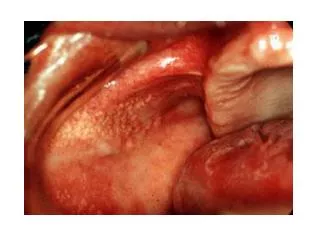
CA Nasopharynx • Carcinoma of the nasopharynx frequently arises from the lateral wall, with a predilection for the fossa of Rosenmuller
LOCAL SPREAD • Anteriorly into the nasal fossa, • Posterolaterally beyond the pharyngobasilar fascia to involve the parapharyngeal and the carotid spaces • Laterally to the pterygoid muscles, • Posteriorly to the prevertebral muscles • inferiorly to the oropharynx
Superiorly, bony erosion of the skull base involving • The floor of the sphenoid sinus, • Clivus • Apex of petrous bone • Basal foramina.
? Intracranially via foramen lacerum • High frequency of perineural spread along the maxillary division (V2) and the mandibular division (V3) of the trigeminal nerve with subsequent intracranial extension through the foramen rotundum and foramen ovale
Nasopharynx Imaging • Before the era of CT - plain radiography • The classic 5 views of NPC that Ho described consist of lateral, submentovertical, occipitosubmental, 25° occipitomental, and occipitomaxillary views
EthmoidSinuses • the Ethmoid air cells • separated from the orbital cavity by a thin, porous bone, the lamina papyracea, and from the anterior cranial fossa by a portion of the frontal bone, the fovea ethmoidalis. • They are in close proximity to the optic nerves laterally and the optic chiasm posteriorly.
The middle ethmoid cells open directly into the middle meatus. • The anterior cells may drain indirectly into the middle meatus via the infundibulum. • The posterior cells open directly into the superior meatus.
The various radiographic positions used to study paranasal sinuses are: • 1. Occipito-mental view (Water's view) • 2. Occipital-frontal view (Caldwel view) • 3. Submento-vertical position (Hirtz position) • 4. Lateral view • 5. Oblique view 39 Degrees oblique (Rhese position)
Maxillary Sinuses • The maxillary sinuses are the largest of the paranasal sinuses. • They are pyramid-shaped cavities located in the maxillae. • The lateral walls of the nasal cavity form the base and the roofs correspond to the orbital floors, which contain the infraorbital canals.
The floors of the maxillary sinuses are composed of the alveolar processes. • The apices extend toward and frequently into the zygomatic bones.
Secretions drain by mucociliary action into the middle meatus via the hiatus semilunaris through an aperture near the roof of the maxillary sinus. • Ohngren'sline is a theoretic plane dividing each maxillary sinus into the suprastructure and infrastructure; it is defined by connecting the medial canthus with the angle of the mandible.