Download
1 / 53
580 likes | 1.69k Views
Magnification of Images. A microscope is an optical instrument that uses a lens or a combination of lenses to magnify and resolve the fine details of an object.The magnified image seen by looking through a lens is known as a virtual image, whereas an image viewed directly is known as a real image.

E N D
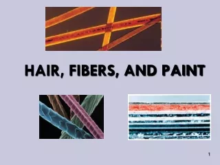
1. The Microscope and Forensic Identification of Hair & Fibers
2. Magnification of Images A microscope is an optical instrument that uses a lens or a combination of lenses to magnify and resolve the fine details of an object.
The magnified image seen by looking through a lens is known as a virtual image, whereas an image viewed directly is known as a real image.
The magnification provided by a lens comes from the process of refraction: light rays bend as they pass through through the lens.
3. Focal Point & Focal Length The point at which parallel rays are converged to an image is the focal point of the lens.
The distance of this point from the lens is the focal length.
4. A Simple Magnifier Object O is placed close to the lens, the rays from it bend but do not intersect.
The observer�s eye follows rays back to the point of apparent origin (I).
The virtual image (I) appears bigger than the object (O)
Moving the lens closer to the eye increases magnification.
5. Terms in Microscopy Magnification:
How much the image is being increased
Depends on the refractive index, curvature and thickness of the lens
Field of View:
Everything that is visible through the eyepiece
When you increase magnification, you decrease field of view
6. Terms in Microscopy Depth of Focus:
The thickness of the specimen that can be seen clearly
Can be used to determine layering of objects
Decreases at higher magnification
7. Types of Microscopes Compound Microscope
Comparison Microscope
Stereoscopic Microscope
Polarizing Microscope
Scanning Electron Microscopes
Microspectrophotometers
8.
Lenses: Ocular and objective (with mirror)
Body tube or head: hollow tube that holds the objective and eyepiece lenses
Stage: platform that supports the specimen
Condenser: focuses light from the illuminator through center of stage Compound Microscope
9. The Compound Microscope Rays from the object (O) pass first through the objective lens forming a real, slightly enlarged, inverted image (I1).
The second lens (eyepiece) acts as a simple magnifier to create an even bigger image (I2).
10. Compound Microscope A compound microscope has at least two lenses:
Objective (lower) lens: produces a magnified and inverted version of the object
Ocular (smaller) lens: produces a virtual image in the viewer�s brain
Magnifying power = power of the objective lens x power of the ocular lens
Working distance = distance between the objective lens and the stage
11. Comparison Microscopes Are used to compare two specimens
Consist of two compound microscopes connected by an optical bridge
Provide a single eyepiece through which the examiner sees both images side by side
Can be lighted from below the stage or via a vertical or reflected illumination system
12. Stereoscopic Microscope
13. Two separate monocular microscopes each with its own set of lenses
Produce a three-dimensional image with a right-side-up, frontward orientation
Offers a large working distance for bulky items
Relatively low magnification (10x-125x)
Can be lighted from below or vertically from above
This microscope is actually two separate monocular microscopes except for the lowest objective lens, which is common to both microscopes
Stereoscopic Microscope
14. Polarizing Microscopes Polarizing microscopes
Include two polarizing filters, a polarizer lens (fixed below the specimen), and an analyzer lens (fixed above the specimen)
The stage with the sample is rotated to determine how the polarized light interacts with the sample
15. Polarized Light
16. Polarizing Microscopes
17. Microspectrophotometers Optical microscopes have been attached to spectrophotometers.
The lamp emits radiation that passed through the sample.
Light is separated according to its wavelength and the spectrum formed is observed with a detector.
18. Infrared (IR) Spectrophotometry Probes molecular vibrations
absorption occurs when the frequency of the IR wave matches the vibrational frequency of a bond in the molecule
Most molecules have numerous vibrations
bond stretching
bond bending
Molecules with different structures have distinctively different IR spectra; therefore it is equivalent to a �fingerprint� of that substance.
19. Example: Photocopier Toner Analysis Analysis must be performed non-destructively
can�t remove toner from paper
physical size of specimen is very small
Use microscope to find sample
Use FT-IR to analyze the toner
20. Scanning Electron Microscope Can magnify 100,000X
Has a depth of focus more than 300X that of an optical microscope
Uses electrons rather than light
Offer much greater resolution than with a light microscope
21. Scanning Electron Microscope
23. Scanning Electron Microscope
24. Hair as Evidence Resists chemical decomposition.
Retains its structural features over long periods of time.
Humans lose about 100 hairs per day so they transfer often and can link suspect, victim and crime scene.
An individual hair cannot result in definitive identification of a person unless it has a DNA tag attached.
25. Collect hair and fiber evidence by using
Wide, transparent sticky tape
Lint roller
Evidence vacuum cleaner
If fibers must be removed from an object
Use clean forceps
fold fiber into a small sheet of paper
store in paper bag
Collecting Hair and Fiber Evidence
26. Collection of Hair Evidence Questioned hairs must be accompanied compared with an adequate number of control samples
from victim
from suspects
From animals
Representative control samples
50 full-length hairs from all areas of scalp
24 full-length pubic hairs
27. The Composition of Hair Hair is composed primarily of keratin, which makes hair resistant to physical change.
Each strand grows out of a follicle.
28. Hair Growth Hair growth stages (Remember ACT):
Anagenic: hair follicle is actively producing the hair; follicle is attached to root (10-1000 days)
Catagenic: transition stage in which the root is pushed out of the follicle (14-21 days)
Telogenic: hair naturally becomes loose and falls out (100 days)
29. The Structure of Hair
Structure of a hair:
Cuticle: scales of hardened, flattened, keratinized tissue which point away from root
Cortex: array of spindle-shaped cells parallel to length of hair embedded with pigment
Medulla: rows of dark-colored cells organized in a pattern specific to the animal species
Cortical fusi: air spaces
30. Cuticle Structure
Coronal Spinous Imbricate
31. Medulla Patterns
32. Forensic Analysis of Hair The following questions apply to hair evidence:
Is the hair human or animal?
Does it match the hair of the suspect?
Does it have a follicle for DNA testing?
33. Is It Animal or Human?
34. Distinguish between animal and human using
Diameter (70-120 micometers)
Pigment distribution (denser toward cuticle)
Cuticle (imbricate)
Medulla (amorphous)
Root shape (bulbous)
Animal vs Human Hairs
35. Assess the hair color, length, and diameter
Examine cuticle, medulla and cortex
Distribution, shape & color intensity of pigment
dyed hair has color in cuticle & cortex
bleaching removes pigment, gives yellow tint
Determine the body area of origin (head, pubic, limbs, face, chest, underarm)
Pests, diseases or contaminants
Can determine presence of drugs by chemical analysis Forensic Analysis of Human Hair
36. Human Hair As Class Evidence Asian
37. Human Hair As Class Evidence Can often determine body area of origin
Can determine shed vs. forcibly removed
Can often determine racial origin
38. Fibers as Evidence Most fibers do not degrade over time.
Fibers are easily transferred from one object or person to another.
Fibers provide evidence of association between a suspect and a crime scene.
Most fiber evidence can only be placed within a class.
39. Fibers are Polymers
40. Identification of Fabrics & Fibers Fiber classification
Chemical identity of fiber and dyes
Type of thread or yarn used
Weave of fabric
Logo or manufacturer identification
41. plants (cotton, hemp, etc.)
animal (wool, silk, etc.)
mineral (asbestos, fiberglass, etc)
man-made (nylon, polyester, etc). Sources of Fibers
42. Classification of Fibers
43. Synthetic Fibers A wide variety of synthetic fibers have replaced natural fibers in fabrics, garments, and rugs.
Synthetic fibers may be made from natural materials that are not normally fiber-like.
There are three types of synthetic fibers:
Cellulose based: produced from cellulose-containing raw materials (rayon)
Synthetic: produced from chemicals made from refined petroleum or natural gas (nylon)
Inorganic: produced from metals, glass or ceramics (fiberglass)
44. Analysis of Fibers Step 1: Natural vs synthetic, use a comparison microscope
Examine the color, diameter, cross-section shape, pitting or striations, etc.
Synthetic fibers have smooth surfaces, uniform size & shape
45. Is It Hair or Fiber? Is it Natural or Synthetic?
46. Analysis of Fibers Step 2: chemical composition of synthetics
Use IR spectroscopy, refractive index or polarized light to identify types of synthetic fiber
Dye can be extracted and the colors separated by thin layer chromatography (TLC)
47. FT-IR Analysis of Fibers
48. Manufacture of Synthetic Fibers Melted or dissolved polymer is forced through fine holes of a spinnerette
Similar to a bathroom showerhead
polymer molecules are aligned parallel to the length of the filament
Shapes of holes in spinneret determine cross-sectional shape of the polymer
49. Fibers: Thread and Yarn Thread and yarn are bundles of fibers woven to create fabrics. Classified as
Filament: continuous length of fiber
Spun: short lengths of fibers that are twisted or spun together
Physical properties of thread and yarn include its texture, number of twists per inch, number of fibers per strand, blend of fibers, color, and pilling characteristics.
50. Woven fabrics consist of intertwining of two sets of yarns that are woven on a loom.
Weave patterns can be used to classify fabrics.
Basic weaves are plain, twill, and satin. Fibers: Thread and Yarn
51. The Microscope and Forensic Identification of Hair & Fibers
52. The Microscope and Forensic Identification of Hair & Fibers
53. The Microscope and Forensic Identification of Hair & Fibers
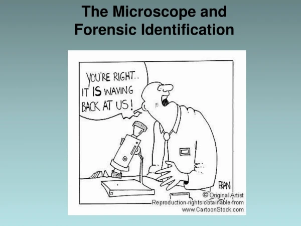
54. Applications of Microscopy: Hair "It's just a formality, but I'll also need a lock of your hair for DNA analysis."