Download
1 / 33
480 likes | 914 Views
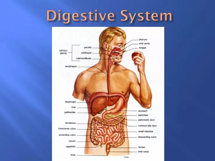
Digestive System. Digestive System – Fig. 15-1, Table 15-1. Digestion – process of changing food substances into forms that can be absorbed; digestive system consists of an alimentary canal & several accessory organs

E N D
Digestive System – Fig. 15-1, Table 15-1 • Digestion – process of changing food substances into forms that can be absorbed; digestive system consists of an alimentary canal & several accessory organs • Digestive structures are adapted for mechanical and chemical digestion; enzyme secretion, and absorption • http://highered.mcgraw-hill.com/sites/0072495855/student_view0/chapter26/animation__organs_of_digestion.html (overview of digestive system)
Digestive System – Fig. 15-1, Table 15-1 • General Characteristics of the Alimentary Canal – various regions are specialized to perform specific functions • Structure of the wall – 4 layers – Fig. 15-2 • Mucous membrane – inner most layer; function to protect, absorption, secretion • Submucosa – functions to nourish surrounding tissues, tranports absorbed materials • Muscular layer (muscularis) – functions in the movement of the tube and its contents; smooth muscle fibers are arranged in circular & longitudinal groups • Serous (serosa) – composed of visceral peritoneum; functions to protect and lubricate
Digestive System • Movements of the tube – Fig. 15-3 • Motor functions include mixing and propelling movements • Mixing – small segments of smooth muscle contraction • Movement by peristalsis – tube undergoes receptive relaxation just ahead of a peristaltic wave
Peristalsis • http://www.youtube.com/watch?v=o18UycWRsaA
Digestive System • Innervation of the tube • Innervated by branches of sympathetic and parasympathetic divisions of the autonomic nervous system • Parasympathetic impulses generally cause an increase in digestive activities; sympathetic impulses generally inhibit digestive activities
Mouth – adapted to receive food and begin preparing it for digestion. It also serves as an organ of speech & sensory reception. Fig. 15-4
Mouth • Cheeks & lips • Cheeks form the lateral walls of the mouth • Lips – highly mobile & possess a variety of sensory receptors useful in judging the characteristics of food • Tongue • Thick, muscular organ that aids in the mastication of food with saliva and moving it toward the pharynx • Rough surface (papillae) aids in handling food & contains taste buds • Lingual tonsils (lymphatic tissue) are located on the root of the tongue
Mouth • Palate • Comprises the roof the mouth and includes hard & soft palates • Soft palate closes the opening to the nasal cavity during swallowing • Palatine tonsils (lymphatic tissue) are located on either side of the tongue in the back of the mouth • Pharyngeal tonsils (adenoids) are located on the posterior wall of the pharynx above the border of the soft palate
Mouth • Teeth • Two sets of teeth develop in sockets of the mandibular & maxillary bones • 20 primary (deciduous) & 32 secondary teeth - Fig. 15-6 • Break food into smaller pieces to increase surface area for digestive enzymes to act upon • Teeth are adapted to handle food in different ways • Incisors – biting • Canines (cuspids) – grasping • Molars - grinding
Mouth • Teeth • Anatomy – Fig. 15-6 – tooth is divided into crown, neck, root, and is attached/anchored by periodontal membrane (ligament) • Enamel – protects • Dentin – makes up the bulk of the tooth
Salivary Glands • Salivary glands – secrete saliva, moistens food, binds food particles together, begins digestion of carbohydrates, makes taste possible, helps cleanse the mouth, & regulates pH in the mouth (pH 6.5 – 7.5) – Fig. 15-7 • Salivary secretions – glands include • Serous cells that secrete digestive enzymes (amylase) that are more liquid/watery • Mucous cells that secrete mucus that is more viscous
Major salivary glands • Parotid – largest; secretion rich in amylase • Submandibular – secretes combinations of enzymes and mucus • Sublingual – secretes mucus
Pharynx & Esophagus – serve as passageways • Pharynx – throat, passage for air & food • Swallowing mechanism – 3 stages • food is mixed with saliva & forces into pharynx • Involuntary reflex actions move the food into esophagus • Esophagus to stomach by peristalsis
Pharynx & Esophagus – serve as passageways • Esophagus • Passes through mediastinum & penetrates diaphragm • Cardiac sphincter – circular ring of muscle that prevents regurgitation • Hiatal hernia – weak diaphragm, some part of stomach, large intestine or other abdominal organ protrudes into thoracic cavity • Heart burn – gastric juice enters esophagus & causes mucosa to become inflamed
Stomach • Mixes food with gastric juices carries on a limited amount of absorption; & moves food to small intestine – Fig. 15-8 • Parts of stomach • Cardiac, fundic body & pyloric regions • Pyloric sphincter – serves as a valve between stomach & small intestine; regulates how much “chyme enters • Gastric glands secrete pepsin (breaks down proteins), HCl (kills bacteria, and keeps stomach at pH of about 2 which is optimal for action of pepsin), and lipase (breaks down lipids)
Stomach • Stomach not well adapted for absorption, however it can absorb some water, lipid soluble drugs, and alcohol • Mixing & Emptying Actions • as stomach fills the walls stretch – internal pressure remains unchanged • Mixing movements (3 directions of smooth muscle) aid in producing chyme; peristaltic waves move chyme to pyloric region • Muscular wall of pyloric region pumps chyme into the small intestine • Rate of emptying depends on fluidity of chyme & the type of food present
Pancreas (accessory organ) – Fig. 15-10 • Pancreatic juice digests all food types • Has a high HCO3- (bicarbonate) content that helps to neutralize chyme & causes intestinal contents to be alkaline • Pancreatic duct merges with bile duct & enters small intestine at duodenum
Liver/Gall Bladder – Accessory Organs • Functions of the liver • Metabolism of fats, sugars, & proteins • Filters blood • Storage of substances – glycogen, iron, vitamins • Detoxifies – converts ammonia to urea • Secretes bile – emulsifies lipids • Structure • Located in upper R. & central portion of abdominal cavity • Highly vascular & divided into lobes • Functional unit – hepatile lobule
Liver/Gall Bladder – Accessory Organs • Function of the Gall Bladder – Fig. 15-10 • Store bile between meals • Release of bile form the common bile duct is controlled by sphincter muscle • Gallstones – cholesterol that has crystallized (pg. 405) • Release of bile is stimulated by cholescystokinin from the small intestine which causes the gall bladder to contract
Small Intestine – extends from pyloric sphincter to the large intestine Fig. 15-9 • Functions • Complete digestion of all nutrients • Absorbs nutrients which then enter blood stream • Transports residues to the large intestine • Secrete digestive enzymes • Parts • Duodenum, jejunum, & ileum • Suspended from the posterior abdominal wall by mesentery (extension of peritoneum)
Small Intestine • Structure (circular folds – plica{e}) • Lined with villi that increase surface area & aid in mixing & absorbing • Microvilli on the free ends of epithelial cells increase surface area even more • Regulation/absorption/movements • Secretion of intestinal enzymes are enhanced by the presence of gastric juice & chime and distension of small intestinal wall • Villi absorb water & nutrients • Movements include mixing by segmentation & peristalsis • Ileocecal valve controls movements of contents from small to large intestine
Large Intestine • Reabsorbs water & electrolytes and forms 7 stores feces – Fig. 15-12, 15-13 • Parts divided into cecum, ascending colon, transverse colon, descending colon, sigmoid colon, rectum, and anal canal • Functions • Little or no digestive function; secretes mucus • Absorption of H2O, electrolytes, & vitamins (produced by bacteria that inhabit large intestine) • Movements – similar to those of small intestine • Feces – stored in rectum and eliminated through anus
Large Intestine • http://www.youtube.com/watch?v=6kg5wZQfADQ (colonoscopy) • http://www.youtube.com/watch?v=nGyzZyxMxkk&NR=1 (colonoscopy showing rectal cancer)