Download
1 / 32
620 likes | 2.86k Views
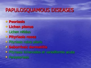
PAPULOSQUAMOUS DISORDERS. Papulosquamous eruptions. Also k/a scaly rash disease Clinical lesions characterized by scaly papules and plaques(may be due to proliferation of cells of epidermis or dermis,infiltration with inflammatory cells or deposits in dermis)

E N D
Papulosquamous eruptions • Also k/a scaly rash disease • Clinical lesions characterized by scaly papules and plaques(may be due to proliferation of cells of epidermis or dermis,infiltration with inflammatory cells or deposits in dermis) • Morphology of papules /plaques varies in different papulosquamouseruption plus there is characteristic scales in each variety.
Causes of sudden scaly rash Common • Eczema Psoriasis • Lichen planus Secondary syphilis • Drug eruptions Less common • PityriasisroseaPityriasisversicolor • Tineacorporis • Exfoliativeerythroderma • Gianotti-Crosti syndrome
History • Common presenting problem- Eruptive scaly rash with or without itching • Differentiation of these diseases are done based upon history and examination
Ask • Rash • Location- Ask where the rash is • Is it at multiple places? • Onset, duration and progression • Associated symptoms Ask if the lesion is: • Itchy– Eczema is extremely itchy • Breathing problems/ chest pain/ joint pain/fever/ red eyes- to R/O other systemic diseases • Aggravating factors • Temperature- Itching of acute eczema increases with rise in temperature • Allievating factors • Ask if anything makes the problem better • Cooler temperature in Acute eczema.
History taking.. • Past Medical History: Previous H/O similar episodes: • Allergic history: • Medical problems in the past: • Hospitalization: • Family History: • Obstetric and Gynecological history: • Sexual History: • Social History:
Mnemonics for history taking: • LIQOR AAA L- Location I - Intensity Q- Quality O- Onset, duration and frequency R- Radiation A- Aggravating factors A- Alleviating factors A- Associated problems • PAM HUGS FOSS P- Previous episodes of C/O A- Allergic History M- Medical problems in the past H- Hospitalization U- Urinary problems G- Gastrointestinal problems S- Sleep Begin with transition question for FOSS F- Family History O- Obstetric and gynecology history S- Sexual H/O S- Social H/O
Examination: • Complete exposure, in bright and uniform light • General examination • Local examination: • Rash - Symmetrical/ asymmetrical - Extensor or flexor - Proximal/ Distal/ Facial - Localised or widespread • Examine scalp, face, eyes, oral mucosa, neck, axilla, nails, groin and joints
Eczema • Eczema literally means ‘to boil out’ • Terms 'eczema' and 'dermatitis' are synonymous. • They refer to distinctive reaction patterns in the skin, which can be either acute or chronic and are due to a number of causes. • It has 2 components: • Clinical • histological
Clinical component 0f THE ECZEMA REACTION Acute • Redness and swelling, usually with ill-defined margins • Papules, vesicles and, more rarely, large blisters • Exudation and cracking • Scaling • Pruritis • Erythema
Histological component Hallmarks- (depending on clinical appearance) spongiosis Hyperkeratosis and acanthosis In the acute stage, oedema of the epidermis (spongiosis) progresses to the formation of intra-epidermal vesicles, which may enlarge and rupture. In the chronic stage there is less oedema and vesiculation but more thickening of the viable epidermis (acanthosis),thickening of stratum corneum(hyperkeratosis) This is accompanied by a variable degree of vasodilatation and T-cell lymphocytic infiltration in the upper dermis.
classification Etiological • Endogenous • Exogenous • combined Morphological • Discoid • Hyperkeratotic • Lichenified • Seborrheic
Endogenous • Atopic dermatitis • Seborrheic dermatitis • Discoid • Pompholyx • Pityriasis alba • Stasis dermatitis • Lichen simplex chronicus Exogenous • Irritant • Allergic • Photo dermatitis • Radiation dermatitis • Infective dermatitis Combined • atopic
Investigations • Acute to be treated before investigations • Chronic: • Patch test • Prick test • Photopatch test • Serological test RAST
Differential diagnosis • Psoriasis • Scabies • Any other papulosquamous lesions
complications Dermatological complications Infecton Dissemination Contact dermatitis Erythroderma Psychosocial complication Anxiety,depression,social comp.
Treatment • General measures Removal of trigger Hydration • Acute phase: topical treatment-soln of either potassium permanganate (0.01%)or aluminium acetate(0.65%)or for large area- calamine lotion /local steroids systemic treatment: short course of steroid , antibiotics,antihistaminics • Chronic phase: nonsteroid-ichthammol,topicalsteroids,pluskeratolytic agents like salicylic acid or urea(for lichenified lesion)antibiotics
Atopic eczema/ dermatitis • Most common form • It is an endogenous eczema triggered by exogenous agents characterised by extremely pruritic recurrent ,symmetric eczematous lesion • Epidemiology: Seen in 3% of all infants , increasing worldwide(decr.breast feeding and increasing pollutants)
Etiology and pathogenesis Associated features:PositiveH/O or Family H/O Asthma, Hay fever, Urticaria or food allergies Genetic predisposition maternal imprinting-that is, they are inherited more often from the mother than from the father Immunological changes: IgElevels,abnormal lymphocytes: Generalized and prolonged hypersensitivity to common environmental antigens Atopic Eczema
Differential diagnosisinfantile seborrheic dermatitisscabiesairborne contact dermatitis
Prevention • EARLY PREVENTION OF ATOPIC ECZEMA 'Restrictions in maternal diet during pregnancy have no effect on the incidence of atopic eczema in an infant at hereditary risk and may adversely affect maternal and/or fetal nutrition. Breastfeeding, however, appears to reduce the prevalence of atopic eczema in early childhood.'
Rx • General measures: Avoid scratching,and avoid triggers Good hydration • Topical therapy Emollients Topical steroids:also with combination of antibiotics /emollients,start with lose dose and if fail then increase the dose,Inlichenified lesion with keratolytic agents like salicylic acid Topical calcineurin inhibitors: immunomodulators-pimecrolimus(1%),tacrolimus(0.03%and 0.1%)
systemic therapy in extensive cases: systemic antibiotics systemic steroids antihistaminicsNew therapiesUVB or PUVA3 mths course of oral evening primrose oil cyclosporin