Download
1 / 31
340 likes | 627 Views
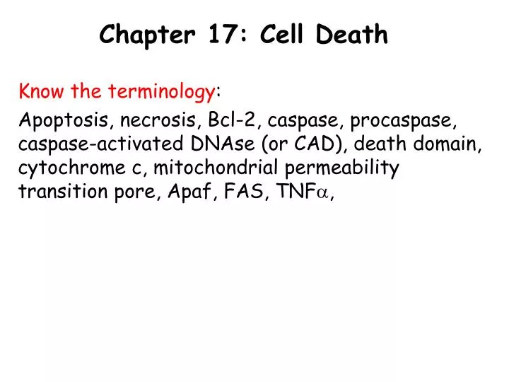
Chapter 17: Cell Death. Know the terminology : Apoptosis, necrosis, Bcl-2, caspase, procaspase, caspase-activated DNAse (or CAD), death domain, cytochrome c, mitochondrial permeability transition pore, Apaf, FAS, TNF a ,. The many forms of cell death. Necrosis:

E N D
Chapter 17: Cell Death Know the terminology: Apoptosis, necrosis, Bcl-2, caspase, procaspase, caspase-activated DNAse (or CAD), death domain, cytochrome c, mitochondrial permeability transition pore, Apaf, FAS, TNFa,
The many forms of cell death Necrosis: Cell death resulting from physical or chemical damage. It progresses in an uncontrolled way and causes local tissue damage or inflammation (in some species) Apoptosis: Controlled form of cell death, where the cell controls its own demise. First, it degrades its internal structure, then dies in a way that is easily handled by local phagocytotic cells.
Why is apoptosis needed? Apoptosis is needed to: 1. Control the death of irreversibly damaged cells, preventing local tissue disruption. For example, -radiation damage -cell cycle defects 2. Remove cells that are unwanted. For example, -morphogenesis (tissue formation) -tissue remodelling
Examples of apoptosis in development Target cells secrete enough “survival factors” to ensure the survival of the appropriate numbers of neurons
Examples of apoptosis in development Embryonic hands/feet start as broad pads, with apoptosis sculpting the final shape by killing the cells between digits
Examples of apoptosis in development Metamorphosis hormones trigger apoptosis of trunk muscles in tail, and induce differentiation of limbs and appendicular muscle
Intracellular events Controlled Cell Death: -cell shrinkage (necrotic cells explode) -cytoskeletal collapse -nuclear envelope disassembles -proteolytic degradation -membrane phospholipid inversion (PS) -membrane display of phagocytotic signals -DNA fragmentation
Caspase activated DNase CAD cuts DNA between histones, resulting in DNA fragments of multiples of 280 bp (a DNA ladder).
DNA Ladder TUNEL Terminal deoxynucleotidyl transferase–mediated dUTP Nick End-Labeling
Meet the executioner: Caspases Caspases are Cysteine-ASPartate proteases: -cysteine in in the enzyme active site -the attack aspartate residues on target -produced by the cell as inactive proenzymes (procaspase) -upon the appropriate signal, the procaspase is cleaved to form the active caspase -who cuts up the procaspase? Another caspase.
Caspase cascade • Self amplifying • Irreversible Initiator caspase (e.g. caspase 9 or caspase 10) Executioner caspase (e.g. caspase 3) Targets
2 routes of apoptotic induction 1. Intracellular route:
Mitochondrial permeability transition pore (MPTP) Recall that the mitochondrial inner membrane has low permeability (it maintains a proton motive force). The outer membrane has porin, which allows small molecular weight molecules to move freely (less than about 7,000 daltons)
Mitochondrial permeability transition pore (MPTP) Cytochrome c (a mobile electron carrier) moves with the intermembrane space. Its too big (~12000 daltons) to cross through porin. When mitochondria completely depolarize, another pore forms from multiple proteins, allowing cytochrome c to escape to cytoplasm. Thus, toxic agents that depolarize mitochondria can trigger apoptosis.
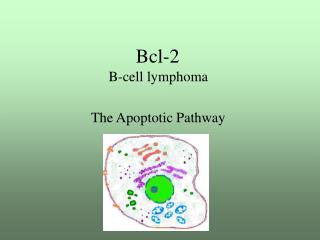
Bcl2 family The mitochondria possess a pore that can allow cytochrome c to escape from intermembrane space The pore opens with massive membrane depolarization Bcl2 (and Bcl XL) are proteins that associate with the pore and keep it closed. Bad and Bid are similar in structure to Bcl2 and bind to anti-apoptotic proteins, blocking their effects.
Bcl2 family form heterodimers to cancel out each others effects
Bcl2 family Bcl2 binds to mitochondria to prevent cytochrome c release Bad binds Bcl2 to prevent it from preventing cell death When Bad is phosphorylated (PKB) it can’t bind Bcl2
2 routes of apoptotic induction 2. Extracellular route:
Extracellular routes of apoptosis Extracellular proteins bind to cell membrane receptors to initiate apoptosis. Include: membrane proteins such as: -FAS ligand (killer T cells) -tumor necrosis factor alpha (TNFa) (macrophages) Activation of death receptor (FAS protein) recruits adaptor proteins with “death domain”
Extracellular routes of apoptosis Adaptor proteins bind (and colocalize) initiator procaspases (e.g. caspase 8). Procaspase 8 has weak proteolytic activity but because they are colocalized, they can attack each other to form an active caspase.
Inhibitors of apoptosis protein (IAPs) Procaspases have some low proteolytic activity that must be held in check in healthy cells Inhibitor of Apoptosis Proteins (IAPs), such as X-linked Inhibitor of Apoptosis (XIAP), act by inhibiting procaspase activity Weak activity Procaspase Very weak activity Procaspase IAP
Inhibitors of apoptosis protein (IAPs) Mitochondria can stimulate apoptosis a second way, by releasing a protein that impairs the effects of IAPs Smac (Second Mitochondrial Activator of apoptosis) Drosophila homologues of Smac include Hid, Grim, Reaper. Smac (and its homologues) stimulate apoptosis by blocking XIAP effects
Procaspase-9 + IAP Procaspase-9 + Smac + IAP Inhibitors of apoptosis protein (IAPs) Procaspase-9 Smac IAP Procaspase-9 IAP Active Inactive
Phosphorylation of Hid Hid can only bind IAP in its dephospho form Phosphorylation of Hid prevents its ability to block IAP’s protective effects
Procaspase-9 + Smac + IAP Procaspase-9 + Smac + IAP Inhibitors of apoptosis protein (IAPs) Procaspase-9 IAP Smac Procaspase-9 Smac IAP Inactive Active
Cancers and apoptosis Cancer is uncontrolled cell growth. Many cancer cells are able to proliferate because they have mutated in a way that prevents the cell from dying. Discussion question: What kind of mutations might disrupt apoptosis?