Download
1 / 24
330 likes | 858 Views
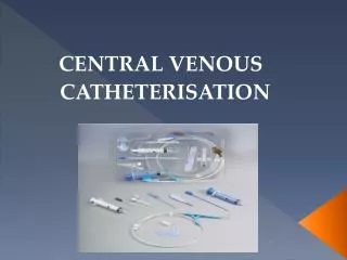
Central Venous Catheterization. UNC Emergency Medicine Medical Student Lecture Series. Objectives . Indications and Contraindications Complications Technique Basic principles Specifics by Site Tips Basic materials. Indications. Central venous pressure monitoring Volume resuscitation

E N D
Central Venous Catheterization UNC Emergency Medicine Medical Student Lecture Series
Objectives • Indications and Contraindications • Complications • Technique • Basic principles • Specifics by Site • Tips • Basic materials
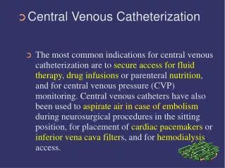
Indications • Central venous pressure monitoring • Volume resuscitation • Cardiac arrest • Lack of peripheral access • Infusion of hyperalimentation • Infusion of concentrated solutions • Placement of transvenous pacemaker • Cardiac catheterization, pulmonary angiography • Hemodialysis
Relative Contraindications • Bleeding disorders • Anticoagulation or thrombolytic therapy • Combative patients • Distorted local anatomy • Cellulitis, burns, severe dermatitis at site • Vasculitis
Complications • Vascular • Air embolus • Arterial puncture • Arteriovenous fistula • Hematoma • Blood clot • Infectious • Sepsis, cellulitis, osteomyelitis, septic arthritis • Miscellaneous • Dysrhythmias • Catheter knotting or malposition • Nerve injury • Pneumothorax, hemothorax, hydrothorax, hemomediastinum • Bowel or bladder perforation
Technique • Seldinger technique • Use introducing needle to locate vein • Wire is threaded through the needle • Needle is removed • Skin and vessel are dilated • Catheter is placed over the wire • Wire is removed • Catheter is secured in place
Basic Principles • Decide if the line is really necessary • Know your anatomy • Be familiar with your equipment • Obtain optimal patient positioning and cooperation • Take your time • Use sterile technique • Always have a hand on your wire • Ask for help • Always aspirate as you advance as you withdraw the needle slowly • Always withdraw the needle to the level of the skin before redirecting the angle • Obtain chest x-ray post line placement and review it
Subclavian Approach • Positioning • Right side preferred • Supine position, head neutral, arm abducted • Trendelenburg (10-15 degrees) • Shoulders neutral with mild retraction • Right side preferred • Needle placement • Junction of middle and medial thirds of clavicle • At the small tubercle in the medial deltopectoral groove • Needle should be parallel to skin • Aim towards the supraclavicular notch and just under the clavicle
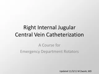
Internal Jugular Approach • Positioning • Right side preferred • Trendelenburg position • Head turned slightly away from side of venipuncture • Needle placement: Central approach • Locate the triangle formed by the clavicle and the sternal and clavicular heads of the SCM muscle • Gently place three fingers of left hand on carotid artery • Place needle at 30 to 40 degrees to the skin, lateral to the carotid artery • Aim toward the ipsilateral nipple under the medial border of the lateral head of the SCM muscle • Vein should be 1-1.5 cm deep, avoid deep probing in the neck
Femoral Approach • Positioning • Supine • Needle placement • Medial to femoral artery • Needle held at 45 degree angle • Skin insertion 2 cm below inguinal ligament • Aim toward umbilicus
Femoral nerve Femoral Vein Femoral artery NAVEL
Post-Catheter Placement • Aspirate blood from each port • Flush with saline or sterile water • Secure catheter with sutures • Cover with sterile dressing (tega-derm) • Obtain chest x-ray for IJ and SC lines • Write a procedure note
Procedure Note • Name of procedure • Indication for procedure • Comment on consent, if applicable • Describe what you did, including prep • Comment on aspiration/flushing of ports • How did patient tolerate procedure • Any complications
Tips • After 3-4 tries, let someone else try • Get chest x-ray after unsuccessful attempt • If attempt at one site fails, try new site on same side to avoid bilateral complications • Halt positive pressure ventilation as the needle penetrates the chest wall in subclavian approach • If you meet resistance while inserting the guide wire, withdraw slightly and rotate the wire and re-advance • Align the bevel with the syringe markings • Use the vein on the same side as the pneumothorax • Withdraw slowly, you will often hit the vein on the way out
Ultrasound-Guided Central Venous Access • Becoming standard of care • Vein is compressible • Vein is not always larger • Vein is accessed under direct visualization • Helpful in patients with difficult anatomy
Compression of vein with US probe Femoral Artery Femoral Vein
References • Clinical Procedures in Emergency Medicine, Roberts and Hedges, 4th edition, 2004 • Clinician’s Pocket Reference, Leonard Gomella, 8th edition, 1997 • Atlas of Human Anatomy, Frank Netter, 2nd edition, 1997