Download
1 / 49
561 likes | 1.04k Views
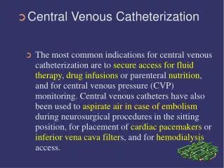
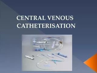
Central Venous Access. Mazen Kherallah, MD, FCCP. Indications. Need for IV access and failure of peripheral access Peripheral access too painful or tenuous Long term IV access anticipated Medications indicated that are toxic to peripheral veins Hemodynamic monitoring

E N D
Central Venous Access Mazen Kherallah, MD, FCCP
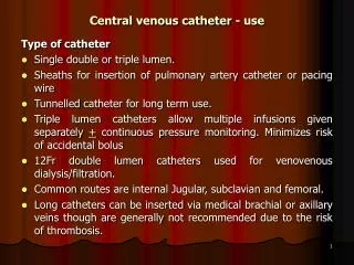
Indications • Need for IV access and failure of peripheral access • Peripheral access too painful or tenuous • Long term IV access anticipated • Medications indicated that are toxic to peripheral veins • Hemodynamic monitoring • Volume resuscitation with large bore central lines • Special procedure: Swan Ganz, dialysis, plasmapheresis
The Procedure • Patient position: • Patient is moved to the side of the bed so physician would not lean over • The bed is high enough so physician would not have to stoop over • Patient should be flat without a pillow, Trendelenburg position if patient is hypovolemic • The head is turned away from the side of the procedure • Wrist restraints if necessary
The Procedure • Skin preparation: • Prepare before putting sterile gloves • Start at the center and work outward the edges • Allow time for the sterilizing agent to dry • Disposable drape under the patient • Betadine or Chlorhexidine are acceptable solution and have activity against gram positive organisms
The Procedure • Drape: • Large enough • Handed sterilely by the assistant • Hole in the area of placement • Prepare the tray: • Handed sterilely by the assistant • Prepare the equipment before starting • Anesthesia • Use local anesthesia with lidocaine
YOUR ROLE AFTER THE INSERTION • Dispose all sharps • Place an occlusive sterile dressing • Flush lumens to maintain patency • Obtain a chest x-ray (ask for order if physician doesn’t mention it) • Monitor site for bleeding • Assess breath sounds • Assess circulation • Assess for hematoma • Document insertion, site, dressing and flushing
USING THE CENTRAL LINE • Flush q shift, before and after use with NS. Some places also require heparin flush • Close clamps when not is use • Check P&P of facility, but usually fluids are changed every 24 hours, tubing changed every 48-72 hours • Dressing is usually changed every 3 days • Line can be used for blood drawing - withdraw and waste 10 cc, then withdraw blood for samples • If port becomes clotted, do not use - sometimes ports can be opened up with urokinase (requires a doctor’s order)
Complications • Immediate • Hemothorax • Pneumothorax • Arterial puncture • Vessel erosion • Nerve Injury • Dysrhythmias • Catheter malplacement • Embolus • Cardiac tamponade
Complications • Delayed • Dysrhythmias • Catheter malplacement • Vessel erosion • Embolus • Cardiac tamponade • Catheter related infection • Thrombosis
Vascular Erosion/Cardiac Tamponade • Large vessel perforation is uncommon • Vessel erosion more common with stiff catheters, like dialysis catheters • Cardiac tomponade occur mainly if the tip is located in the RA • Complication is fatal in 2/3 of cases
Air Embolism • Air is sucked in through the catheter due to negative intrathoracic pressure during inspiration • Air can be pushed with flushing the catheter if it was not pulled back before flushing • Complication is uncommon but can be fatal • Manifests with hypoxemia, cardiovascular collapse, mental status changes and livedo reticularis • Place patient to left lateral position if suspected
Bleeding • More common in patients with coagulopathy • Easily controlled with femoral or IJ sites • Place local pressure and correct coagulopathy
Arterial Puncture and Cannulation • If the artery is puncture local pressure is applied for 3-5 minutes, observe for hematoma formation • If the artery is cannulated, pulsatile reflux of blood can be noticed, blood gas analysis reveals arterial. • The catheter should not be used, and remove it after coagulopathy is corrected if present
Thrombosis • Sleeve fibrin surrounding the catheter (occurs on the majority of catheters) • Mural thrombus on the wall of the vein (10-30% of catheters) • Occlusive thrombus (1-10%)
Pneumothorax • Most likely, pneumothorax is noticed after CXR is seen, unless patient developed tension pneumothorax with hypoxemia, cardiopulmonary collapse and absent breath sound • Small pneumothorax may be watched closely without chest tube placement in the spontaneously breathing patients • Large pneumothorax requires chest tube placement • Even small pneumothorax in patients on positive pressure ventilation requires chest tube placement
Catheter-Related Sepsis • Late complications • Femoral > IJ > subclavian • Triple lumen > single lumen • Large bore > smaller catheter • Sterility of procedure • Number of hub manibulations
Reading the mean of an A wave 22+10/2=16
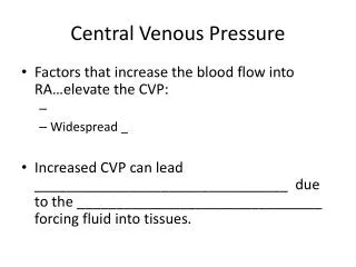
Increased CVP: Right heart failure Right myocardial infarction Cardiac tomponade Tricuspid insufficiency Left to right shunt Pulmonary emboli COPD and cor pulmonale ARDS Excess fluid Tricuspid stenosis Decreased CVP hypovolumia Decreased venous return Excessive veno or vasodilation Shock Hemodynamic MonitoringCentral Venous Pressure: normal 4-10
Central Venous Pressure Tracings • Normal EKG tracing and right atrial pressure waveform • Atrial fibrillation • Atrioventricular dissociation
Central Venous Pressure Tracings • Normal EKG and right atrial waveforms • Tricuspid stenosis • Mild to moderate tricuspid insufficiency • Severe tricuspid insufficiency • Constrictive pericarditis
Large A wave Secondary to Loss of Atrioventricular SynchronySimultaneous Atrial and Ventricular Contraction