Download
1 / 21
210 likes | 253 Views
CHAPTER 3 EQUIPMENT OPERATION AND QUALITY CONTROL. EXAMINATION REVIEW FOR RADIOGRAPHY. ARRT Certification Exam: Equipment Operation and Quality Control. Twenty-two questions (11% of the examination) Three primary sections Principles of Radiation Physics Imaging Equipment

E N D
CHAPTER 3EQUIPMENT OPERATION AND QUALITY CONTROL EXAMINATION REVIEW FOR RADIOGRAPHY
ARRT Certification Exam: Equipment Operation and Quality Control • Twenty-two questions (11% of the examination) • Three primary sections • Principles of Radiation Physics • Imaging Equipment • Quality Control of Imaging Equipment and Accessories
Principles of Radiation Physics: X-ray Production • Primary requirements • Source of free electrons • Cathode (− charge) • Heated filament • Focusing of electrons • Focusing cup • Acceleration of electrons • Applied kVp • Deceleration of electrons • Rotating tungsten anode (+ charge)
Principles of Radiation Physics: X-ray Production—(cont.) • The x-ray tube produces x-rays due to the conversion of high-speed electrons (kinetic energy) into electromagnetic energy (x-ray photons, heat, and light). • X-ray tube efficiency: 1% x-ray production • 99% of energy produced: heat • Two primary atomic interactions occur in the x-ray tube • Bremsstrahlung (85% of radiation produced) • Characteristic (15% of radiation produced)
Bremsstrahlung Radiation • Projectile electron loses kinetic energy (KE) as it interacts with the nuclear field of a target atom. • Deceleration occurs due to a change in direction of the projectile electron. • Lost KE converted to x-ray photons • Produces varying energy levels and wavelengths of x-ray photons
Characteristic Radiation • Projectile electron ionizes a k-shell electron of a target atom. • Opening in K-orbit replaced by an outer shell electron (transition) • Characteristic radiation photon released • Produces fixed energy x-ray photons • In a tungsten target, no characteristic radiation will be produced under 69 kVp.
Fundamental Properties of Electromagnetic Radiation and X-ray Photons • Descriptive or controlling factors • Frequency • Wavelength • Velocity • Energy • Inverse square law
Characteristics of the X-ray Beam • Descriptive or controlling factors • Quality • Voltage • Quantity • Current • Filtration • Target material • Voltage waveforms/generators
Primary versus Remnant (Exit) Radiation • Primary radiation • Primary beam • Leakage radiation • Remnant or exit radiation
Imaging Equipment: Operating Console • Power: (on/off) • kVp control • Provides E for beam penetration • mA control • Controls quantity or intensity of x-ray beam • Timer • Used in conjunction with mA to control the intensity of x-ray beam
Imaging Equipment: Automatic Exposure Control • Components • Ion chambers or sensors • Density controls • −2 to +2 • Backup timer
Imaging Equipment: Circuit Components • X-ray tube • Produces x-rays • Transformers • Controls and varies voltage • Incoming current: 60 Hertz AC • Each peak = one pulse • Rectifers • Converts AC to DC • Generators • Single phase • Three phase
Imaging Equipment: Accessory Devices • Beam restriction devices • Adjustable collimator • Aperture diaphragm • Cones and cylinders • Grids • Stationary • Reciprocating (Bucky device)
Dedicated X-ray Equipment • Chest: freestanding system for upright imaging • Often incorporates • Automatic vertical tracking • Automatic collimation • Tomography: focuses on a single plane • Tube and IR move synchronously around a fulcrum
Fluoroscopic Equipment • Used for real-time imaging • Conventional and digital systems available • Fixed or mobile (C-arm) systems • Components • X-ray tube • Image intensifier • Automatic brightness control • Display screen/monitor • Recording device(s)
Digital Imaging • Filmless equipment • Two types • Computed radiography (CR) • Digital radiography (DR)
Digital Imaging: Computed Radiography (CR) • Components and image acquisition • Reusable imaging plate • Photostimulable phosphor (PSP) • Stores latent image • Laser reader • Stimulates phosphor to release light photons • Monitor • Displays image • Fluorescent light • Erases PSP
Digital Imaging: Digital Radiography (DR) • Components and image acquisition (two types) • Direct panel DR (amorphous selenium) • Converts x-ray photons directly to an electric signal, sent to a computer • Indirect panel DR (amorphous silicon) • Converts x-ray photons to light to an electric signal, sent to a computer
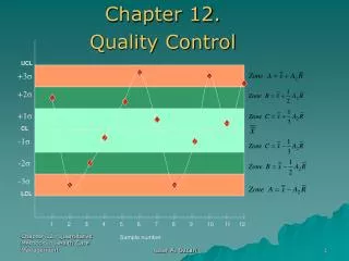
Quality Control (QC) of Equipment and Accessory Devices • Beam restriction devices QC tests • X-ray/light field alignment: ±2% of SID • Central ray alignment • Shielding QC tests (aprons, gloves, and gonadal shields) • Visual inspection for tears or cracks • Fluoroscopy check for holes or cracks
Digital Imaging Quality Control: Artifacts • Types/causes • Debris/dust • Scratches • Incomplete erasure • Ghost images • Fog • Software
Digital Imaging: Monitor Quality Control • Checked monthly or quarterly • SMPTE (Society of Motion Picture and Television Engineers) test pattern • Tests monitor for • Sharpness • Distortion • Luminance