Download
1 / 70
1k likes | 2.32k Views
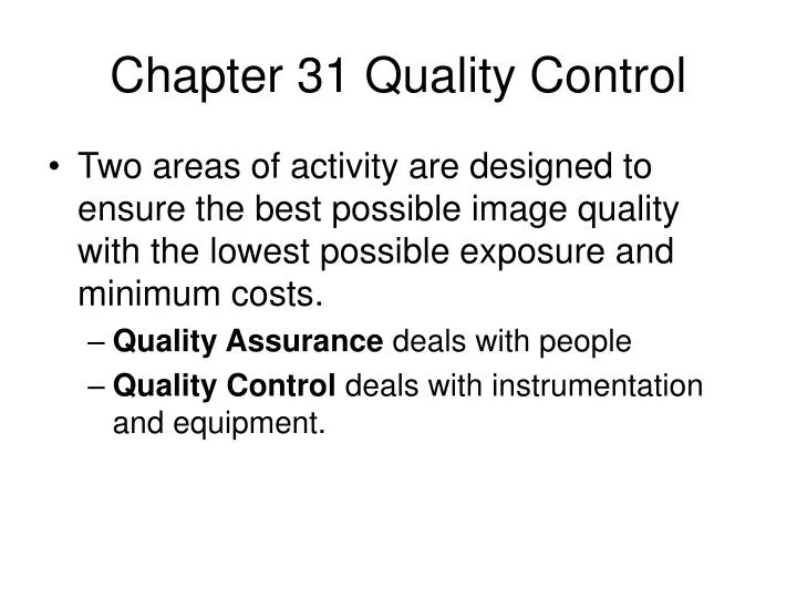
Chapter 31 Quality Control. Two areas of activity are designed to ensure the best possible image quality with the lowest possible exposure and minimum costs. Quality Assurance deals with people Quality Control deals with instrumentation and equipment. Ten Step Approach to Quality Assurance.

E N D
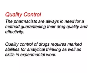
Chapter 31 Quality Control • Two areas of activity are designed to ensure the best possible image quality with the lowest possible exposure and minimum costs. • Quality Assurance deals with people • Quality Control deals with instrumentation and equipment.
Ten Step Approach to Quality Assurance • Assign responsibility • Delineate scope of care • Identify aspects of care • Identify outcomes that effect the aspects of care. • Establish limits of the scope of assessment.
Ten Step Approach to Quality Assurance • Collect and organize data. • Evaluate care when outcomes are reached. • Take action to improve care • Assess and document actions • Communicate information to organization-wide QA Program
QA Projects • Things that QA can evaluate includes • Scheduling of patients • Instructions given to patients • Wait times in the office • Interpretation of films • Retake analysis • Record accuracy
QA Program • Quality Assurance deals with people and processes used to complete tasks. • QA involves training and record keeping. • As the owner of the equipment, you will be responsible for your radiology services. • The State of California Department of Radiologic Health established the Standards of Good Practice that is the foundation of QA and QC in radiography.
QA and QC Requirements • Degree of requirements vary by state. California and New York have very tight standards for quality control of the radiographic and processing equipment. • We are required by statue to teach QA and QC in the radiology program. It is covered in detail in 9th Quarter. My textboook covers QC in detail.
Quality Control • An acceptable QC program has three steps: • Acceptance Testing • Routine performance monitoring • Maintenance
Acceptance Testing • The x-ray machine, cassettes and film processor or digital system are the largest capital expense you may experience. • It makes economic sense to make sure that the equipment meets the performance standards. • It is recommended that a third party such as a health physicist do the testing.
Acceptance Testing • Areas that should be tested include on the x-ray machine: • Shielding of Room • Focal spot size • Calibration of mA, timer or mAs • Calibration of kVp • Linearity of exposure • Beam alignment • Grid centering • Collimation • Filtration (HVL)
Acceptance Testing • Areas that should be tested on the x-ray cassettes: • Screen contact • Screen speed • Light leaks • Light spectrum matching
Acceptance Testing • Areas that should be tested on the x-ray film processor: • Developer temperature • Replenishment rates • Travel time • Water flow • Hypo retention
Quality Control • The acceptance testing ensures that the machine was installed and calibrated properly. • The performance may drift or deteriorate over time. Consequently, periodic testing is required to monitor the performance. • With the exception of film processing most testing is annual or semiannual.
Quality Control • After a major repair, the machine should be retested to ensure that it was repaired properly. • When the testing shows that the machine is not performing properly, service or preventive maintenance is required. • Manufactures establish recommended preventive service schedules. When these are followed many repairs become unnecessary.
Radiographic Quality Control • Areas of concern in x-ray machine • Focal Spot Size will impact spatial resolution • Filtration will impact patient exposure • Collimation will impact patient exposure • kVp calibration will impact image quality and exposure. • Exposure timer accuracy will impact image quality and exposure
Radiographic Quality Control • Areas of concern in x-ray machine • Exposure linearity will impact exposure and image quality • Exposure reproducibility will impact exposure and image quality. • Alignment of tube and image receptor will impact exposure and image quality.
Focal Spot Testing • When the machine is installed or the tube is replaced, the focal spot size should be measured. Then annually thereafter. • A pin hole camera, star test pattern or line pair test tool. • As the tube ages, the focal spot tends to grow and spatial resolution is lost.
Filtration • The filtration is measured but determining the half value layer of the beam at specific exposure levels. Minimum filtration is 2.5mm aluminum. • As a tube ages, tungsten will plate the x-ray port and increase filtration. This can cause technique problems. Inadequate filtration will significantly increase patient exposure.
Collimation • If the collimation is misaligned, intended anatomy can be missed. • It can be tested in many ways from using pennies to using test patterns. • Misalignment can not exceed ± 2% of the SID. • It is tested semiannually and after the replacement of the collimator lamp.
kVp Calibration • In diagnostic radiology, any change will impact patient exposure. A variation of about 3% will impact contrast and image density. • Can be tested with filtered ion chambers, filtered photodiodes or even a cassette with calibrated filters. • Tested annually.
Exposure Timer Accuracy • The exposure time is the responsibility of the operator. It will impact the density of the image and spatial resolution. • Tested with an ion chamber, multi-meter internally or even a spinning top. • Exposure time must be within ±5% for exposure times greater than 10 ms and ±20% less than 10 ms.
Exposure Linearity • Many combinations of mA and time will produce the same mAs value. The ability of the machine to produce a constant level of exposure with various combinations of mA and time is called exposure linearity. • Can be tested with a step wedge and densitometer or rate meter. • Should be within 10% for adjacent stations.
Exposure Reproducibility • Any exposure using the same factors should produce the same level of density and contrast on the image. • Sequential exposure should be reproducible to within ± 5% • Can be tested with a rate meter or step wedge and densitometer.
Darkroom and Processing • The development of the image is dependent upon the temperature of the developer, it’s concentration and how long the film is in the developer. • The film is sensitive to variations in the environment and processing from the time it is manufactured until it is processed. • Darkroom and Processor QC is the key process of Quality Control.
Processing • Processor densitometry is performed daily before the first patient is exposed. • A sensitometer is used to produce a step wedge image on the film that is evaluated with a densitometer. • The densitometer reads the optical density of the processed image. • A digital thermometer is used to test the chemical temperatures in the processor.
Processing • Key densities on the processed film are measured and then graphed. • Base plus Fog is measured on an area of unexposed film to check the darkroom environment. • Speed is tested at the level of exposure that produces a density of 1.25OD • Contrast is tested at the level that produced a density of 0.40 OD and one that produced a density of 2.20.
Processing • By monitoring these densities, problems with film processing can be detected before image quality deteriorates. • In 9th Quarter we will cover how to perform processor QC and problem solve.
Waste Records • Since used fixer is classified as a hazardous waste material, it is important to maintain accurate records of usage and disposal. • The extent of records vary by city, county and state. You are responsible for the proper disposal of the waste. Some regions include developer as hazardous waste.
Silver Recovery • If the silver ions are removed from the fixer, it may be disposed of in the normal waste when diluted with water. • There are two primary types of silver recovery systems. • Metallic replacement uses steel wool and can recover 95% of the silver in the effluent • Electrolytic recovery passes direct current through the solution and nearly pure metallic silver is deposited on the cathode.
Silver Recovery • Old radiographic films and repeated films are retained for silver recovery. X-ray images can not be disposed of in normal trash. • They also can not be used to clean the processor rollers. • Waste recovery companies will either burn or chemically remove the silver from the film.
Performance standards for film processor and darkroom equipment
Accessory QC • The cassettes and screens are the area of chief concern. Problems with either will result in artifacts on the images and increased retakes. • The screens need to be properly cleaned frequently. Mammography screens are cleaned daily. California recommends monthly cleaning.
Accessory QC • Dirty screens produce white artifacts on the image. • Multiple white artifacts indicate the need to replace the screens. • Screen contact is tested semiannually. A problem with screens contact will cause a loss of resolution. • As screens age, they loose speed so this is also tested.
Accessory QC • The other important accessory is the gonad protection devices and lead aprons. • Improper care of the apron can result in cracks and holes in the lead that reduces their effectiveness. • Aprons and shields are tested semiannually. The easiest was to test them is with video-fluoroscopy but film can be used.
Record keeping • All electromechanical devices need periodic service. • There are three types of maintenance. • Scheduled maintenance such as processor monthly or weekly service. It includes observing moving parts and lubrication. • Preventive maintenance is planned service and replacement of parts at regular intervals before they fail at inopportune times.
Record keeping • Non-scheduled maintenance is the worst type of service because it impacts patient service. It may also be very expensive. • With proper scheduled and preventive maintenance, non-scheduled service can be minimized. • All service schedules should meet manufacture recommendations. • All service should be documented as part of the quality assurance program.
Retake Analysis • Required part of a QA program in California. • Evaluation includes • View repeated • Cause of the repeat • Rate of retakes should be less than 5%. • Information can be gathered from the log that the state mandates for patients being exposed to radiation.
Retake Analysis • Done every three months using a relatively large sample of data to see trends in: • Type of examination being repeated. • Reasons for the repeated films. • Determine if additional training or review is needed. • Determine if equipment service might be required.
Repeat Analysis • Your patient log can be designed to capture both films usage and repeated films. • Data is gathered from log for analysis • Repeated films can be put into two main categories: • X-ray Personnel Errors • Equipment malfunctions
X-ray Personnel Errors • Failure to measure patient. • Use of improper technical factors (mAs, kVp or distance) • Incorrect positioning • Improper Collimation • Improper use of accessories such as cassettes, grids or filters
X-ray Personnel Errors • Improper handling of exposed or unexposed films. • Failure to clearly communicate to the patient breathing instructions and to remain still. • Failure to observe patient during exposure.
X-ray Equipment and Accessory Failure or Malfunctions • Inaccurate calibration of kVp and mA. • Inaccurate timer calibration • Dirty or damaged cassettes • Improperly labeled or damaged grids • Malfunctioning collimator • Improper film storage or processor function.
Reasons for Retake Films • Over or under exposure accounts for over 50% of retakes nationally. • Errors in positioning (25%) • Patient motion 11% • Processing errors 6% • Wrong view, beam alignment,cassette screen or grid errors and artifacts.
Retake Films by Region • Cervical Spine Exams = 7% • Thoracic Spine Exams = 17% • Lumbar and Abdominal X-rays = 40% • Skull, Chest,Lower Extremities = 15% • Majority or retake results in unnecessary exposure to gonads or blood forming organs of the body.
Daily Log Design • Most regulatory agencies will require a log of patients having radiation exposure. • Columns can be added to capture film usage, repeats, views repeated and reason. • This data can be gathered and analyzed.
Retake Analysis • In this example, most of the retakes were of the T-spine. Potential reasons include: • Improper use of filters • Incorrect measurements • Faulty technique chart
Retake Analysis • The reason of each retake is recorded and percentages are computed to determine the overall rate and rate by reason. • Less than 5% is ideal.
Quality Assurance and Digital Radiography Information gathered for this lecture came from American College of Radiology Practice Guidelines for Digital Radiography 10/10/07 “Quality Management in The Imaging Sciences “ Jeffrey Pap Mosby 1998