Download
1 / 46
460 likes | 463 Views
This study explores the generation mechanisms and distinguishing features of physiological sharp wave ripples and epileptiform bursts in the hippocampal CA3 area. Experiments are conducted on thick hippocampal slices using an in vitro approach that allows for parallel measurement of network dynamics and neuronal activity. The goal is to understand the quantitative differences and transitions between physiological and pathological network activities.

E N D
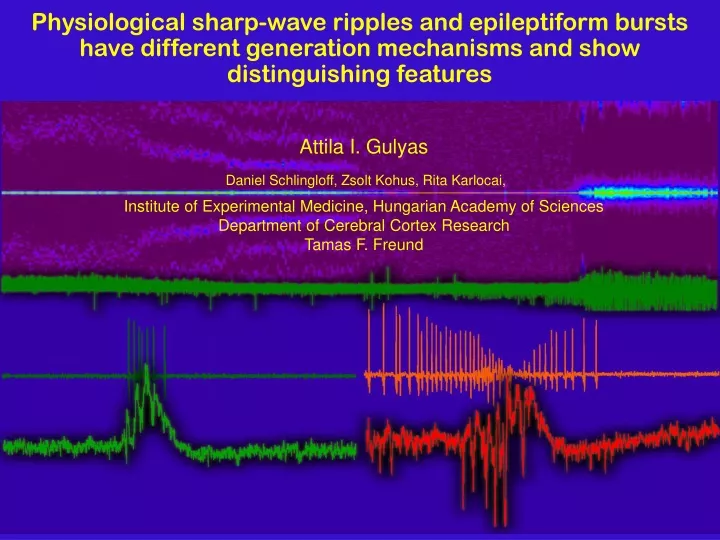
Physiological sharp-wave ripples and epileptiform bursts have different generation mechanisms and show distinguishing features Attila I. Gulyas Daniel Schlingloff, Zsolt Kohus, Rita Karlocai, Institute of Experimental Medicine, Hungarian Academy of Sciences Department of Cerebral Cortex Research Tamas F. Freund
Our laboratory studies how the interaction of excitatory and inhibitory neurons result in different types of network activities. Questions: How and why the hippocampal CA3 area generates different types of physiological and pathological network dynamics? What and why goes wrong in the pathological state? Method: • thick (450-600um) hippocampal slices in modified submerged chamber (4-8ml/min). We use an improved in vitro hippocampal slice preparation (CA3 area) that generates in vivo observed, high activity levels and different behavior associated activity types. Record at 10kHz with patch pipette.
The slices can produce: • gamma oscillation that is present in the hippocampus during exploratory behavior and SWS and is embedded into theta • SWRs, that are essential in memory consolidation during still alertness and REM • and epileptiform (EPI) events, that occur in pathological conditions • In vivo theta embedded gamma, sharp wave ripple and epileptic seizure • In vitro produced gamma, SWR and seizure like event Timofeev et al 2004 Klausberger et al 2003 • the in vitro approach allows: • parallel measurement of LFP (network dynamics) as well as single or dual loose-patch or whole cell measurement of neuronal activity and inputs from identified neuron subpopulations • quick and reversible modulation of network activity • allow local drug application to selectively manipulate components of the network without influencing general dynamics • real experimentation to establish causality
The questions I will try to answer: • What are the quantitative differences between in vitro SWRs and epileptiformbursts (~inter-ictal events, IIEs)? • How are SWRs generated? • How and why physiological synchronous events (SWRs) turn into pathologic epileptiform bursts (~IIEs) in the high K+(and in other [zero Mg2+, 4AP, GABAa blockade]) model? • How and why can the same network generate different activities with distinct dynamics? • What is the difference between physiological and pathological High-Frequency Oscillations (pHFO)? • Why pHFOs indicate sites of impaired inhibition?
Q: What are the quantitative differences between in vitro SWRs and epileptiform bursts (~inter-ictal events, IIEs)?
SWRs and epileptiform bursts (EPI) are different transient high activity events Karlocai 2014 Brain • Recording LFP, extracting dynamics, unit activity and unit frequency • we evoked SWR to epileptiform activity transitions in several ways (high K, 8.5mM, partial GABAa block, 4AP, zero Mg) • EPIs and SWRs have different parameters (amplitude, duration, HFO frequency, unit firing rates) • EPIs are separated from SWRs by a disorganized transitory period in all models • synchrony of cellular activity first falls apart and following an asynchronous period gradually reorganizes into an other type of synchrony
SWRs and epileptiform bursts (EPI) are different transient high activity events Karlocai 2014 Brain • Recording LFP, extracting dynamics, unit activity and unit frequency • we evoked SWR to epileptiform activity transitions in several ways (high K, partial GABAa block, 4AP, zero Mg) • EPIs and SWRs have different parameters (amplitude, duration, HFO frequency, unit firing rates) • EPIs are separated from SWRs by a disorganized transitory period in all models • synchrony of cellular activity first falls apart and following an asynchronous period gradually reorganizes into an other type of synchrony
SWRs and epileptiform bursts (EPI) are different transient high activity events Karlocai 2014 Brain • Recording LFP, extracting dynamics, unit activity and unit frequency • we evoked SWR to epileptiform activity transitions in several ways (high K, partial GABAa block, 4AP, zero Mg) • EPIs and SWRs have different parameters (amplitude, duration, HFO frequency, unit firing rates) • EPIs are separated from SWRs by a disorganized transitory period in all models • synchrony of cellular activity first falls apart and following an asynchronous period gradually reorganizes into an other type of synchrony
SWRs and epileptiform bursts (EPI) are different transient high activity events Karlocai 2014 Brain • Recording LFP, extracting dynamics, unit activity and unit frequency • we evoked SWR to epileptiform activity transitions in several ways (high K, partial GABAa block, 4AP, zero Mg) • EPIs and SWRs have different parameters (amplitude, duration, HFO frequency, unit firing rates) • EPIs are separated from SWRs by a disorganized transitory period in all models • synchrony of cellular activity first falls apart and following an asynchronous period gradually reorganizes into an other type of synchrony
Q: How are sharp waves generated? What initiates a SWR? How is ripple oscillation (nHFO) generated?
Cell types show characteristic activity pattern and inputs during SWRs Hájos 2013 JNeurosci
How are SWRs initiated, shaped and terminated? Hájos 2013 JNeurosci • We analyzed the behavior of identified neurons in the CA3 area during in vitro : • physiological SWRs are short transient bursts of activity • between 2 SWRs PCs are active at a low but steady level • while inhibitory neurons are silent • ~50msec before a SPW peak activity starts to grow exponentially • after the exponential phase E and I activity is ripple frequency modulated Schlingloff 2014 JNeurosci
SWR initiation is stochastic • SWR IEI distribution follows gamma distribution • changing excitability and network size shifts SWR rate according to the predictions of the stochastic model of recurrent burst initiation with a refractory period • SWRs are initiated by the combination of a stochastic and a refractory mechanism: when sufficient number of CA3 PCs are active at the same time activity starts to build up in their recurrent collateral system exponentially, the burst is followed by a silent period
Perisomatic inhibition is current and rhythm generator in str. pyramidale Local drug puff, influencing local integration but not global dynamics revealed: • perisomatic inhibition generates LFP in str.pyramidale • phase locking of cellular activity is lost when perisomatic inhibition is removed
Ripple oscillation is generated by PV-positive neurons cell type specific blockade of inhibitory transmission revealed: • PV but not CCK containing perisomatic neurons are responsible for ripple activity and field potential generation • PVBCs are current as well as pattern generators and phase lock unit firing
Ripple oscillation is generated by PV-positive neurons • transient optogenetic driving of PV-positive cells evokes SWRs • when excitatory transmission is blocked optical stimulation evokes ripple-like events
Ripple oscillation is generated by PV-positive neurons • silencing PV cells blocks SWRs
Ripple oscillation is generated by PV-positive neurons • dual recording of SWRs with two nearby electrode in CA3 • small vertical cut on the border of str. pyramidale and str. lucidum, that severs horizontally running PV+BC main axons
Ripple oscillation is generated by PV-positive neurons local disconnection of subnetworks revealed: • reciprocal inhibition among PV-positive cells is essential for synchronized ripple oscillation • PVBCs are current as well as pattern generators
Ripple oscillation is generated by PV-positive neurons local disconnection of subnetworks revealed: • reciprocal inhibition among PV-positive cells is essential for synchronized ripple oscillation • PVBCs are current as well as pattern generators local inactivation of inhibition revealed: • reciprocal inhibition among PV cells is required for phase locking and rhythm generation
How are SWRs initiated and generated? 4 stages of SWR evolution • between 2 SWRs PCs are active at a low but steady level, while inhibitory neurons are silent • SWRs are ininitiated stochastically through a Poisson process, when suprathreshold amount of pyramidal cells fire together • this will initiate an exponential buildupof activity in the recurrent PC network(proposed by Menendez de La Prida and Miles for EPI initialisation) Schlingloff 2014 JNeurosci
How are SWRs initiated and generated? • PV positive BCs start to be recruited and synchronize neuronal activity at ripple frequency (via the FINO mechanism), this results in the physiological HFO shaped primarily by IPSPs • SWRs can beterminated by the combinaton of several refractory mechanisms (slow inhibition, short-term plasticity, PC accomodation) Schlingloff 2014 JNeurosci
Transition to pathology 1 What goes wrong at the cellular level?
Changes in cellular and network parameters underlie pathological activity • We measured cellular paramters and synaptic transmission in high K+: • excitability increases • perisomatic and dendritic IPSC amplitude decreases • EPSC amplitude increases • due to strong Short-Term Depression of PVBC IPSCs perisomatic inhibitory transmission fails
Changes in cellular and network parameters underlie pathological activity • We measured cellular paramters and synaptic transmission in high K+: • excitability increases • perisomatic and dendritic IPSC amplitude decreases • EPSC amplitude increases • due to strong Short-Term Depression of PVBC IPSCs perisomatic inhibitory transmission fails
Changes in cellular and network parameters underlie pathological activity • We measured cellular paramters and synaptic transmission in high K+: • excitability increases • perisomatic and dendritic IPSC amplitude decreases • EPSC amplitude increases • due to strong Short-Term Depression of PVBC IPSCs perisomatic inhibitory transmission fails
Changes in cellular and network parameters underlie pathological activity • We measured cellular paramters and synaptic transmission in high K+: • excitability increases • perisomatic and dendritic IPSC amplitude decreases • EPSC amplitude increases • due to strong Short-Term Depression of PVBC IPSCs perisomatic inhibitory transmission fails
Transition to pathology 2 What are the consequences? What goes wrong on the network (dynamics) level?
PVBCs get into depolarization block during EPIs and pyramidal cell firing becomes uncontrolled • Parallel recording of LFP and cellular activity • all examined cell types fire differently during EPI than during SWRs • cell increase their firing rate: all PCs fire repetitively during EPIs • INs fire more during EPIs than during SWRs • PV positive basket cells stop firing at the summit of the event, • their AP amplitude decreases before the cessation of firing
PVBCs get into depolarization block during EPIs and pyramidal cell firing becomes uncontrolled • Parallel recording of LFP and cellular activity • all examined cell types fire differently during EPI than during SWRs • cell increase their firing rate: all PCs fire repetitively during EPIs • INs fire more during EPIs than during SWRs • PV positive basket cells stop firing at the summit of the event, • their AP amplitude decreases before the cessation of firing
PVBCs get into depolarization block during EPIs and pyramidal cell firing becomes uncontrolled • Parallel recording of LFP and cellular activity • all examined cell types fire differently during EPI than during SWRs • cell increase their firing rate: all PCs fire repetitively during EPIs • INs fire more during EPIs than during SWRs • PV positive basket cells stop firing at the summit of the event, • their AP amplitude decreases before the cessation of firing
PVBCs get into depolarization block during EPIs and pyramidal cell firing becomes uncontrolled • Parallel recording of LFP and cellular activity • cells generate the observed firing pattern when held at -40mV meaning that cells are depolarized to this potential by the high K • membrane potential matches theoretical membrane potential calculations for high K ACSF • at the peak of the EPI event PV-positive cells get into depolarization block and stop firing, because they receive the strongest excitatory input among hippocampal INs
PVBCs get into depolarization block during EPIs and pyramidal cell firing becomes uncontrolled • Parallel recording of LFP and cellular activity • cells generate the observed firing pattern when held at -40mV meaning that cells are depolarized to this potential by the high K • membrane potential matches theoretical membrane potential calculations for high K ACSF • at the peak of the EPI event PV-positive cells get into depolarization block and stop firing, because they receive the strongest excitatory input among hippocampal INs
PVBCs get into depolarization block during EPIs and pyramidal cell firing becomes uncontrolled • Parallel recording of LFP and cellular activity • cells generate the observed firing pattern when held at -40mV meaning that cells are depolarized to this potential by the high K • membrane potential matches theoretical membrane potential calculations for high K ACSF • at the peak of the EPI event PV-positive cells get into depolarization block and stop firing, because they receive the strongest excitatory input among hippocampal INs
How are EPIs initiated, shaped and terminated? A buildup of activity is spontaneously initiated in a subpopulation of PCs PVBCs are recruited, but due to pathologically strong excitation they get into depolarization block, as well as their transmission fails (see Andrew Trevelyan and also Sydney Cash/ OmarAhmed) PC start uncontrolled pseudo-synchronous burst firing that is manifested as a pHFO, accommodation of PC firing results in decreased E drive, so that PVBCs can recover from depol block and can stop the event • Difference from SWRs • during EPIs control is lost for several reasons: • the balance of excitation/inhibition and cellular excitability changes • PVBCs get into depolarization block • their inhibitory transmission fails (strong STD) Karlocai 2014 Brain
Differences in the HFOs of physiological and pathological synchronous events What are the differences? What are the underlying mechanisms? EPI SWR • pHFOs during EPIs have higher frequency (300-400Hz) than SWR nHFOs (160-200Hz) (see again Menendez de lla Prida ) • during the pHFO the frequency and power drops (use wavelet!)
HFOs are generated by different mechanisms in SWRs and EPIs LFP recording during SWRs and EPIs dual local puff of TTX or GABAzine during both states to inactivate AP generation and perisomatic inhibition, respectively
HFOs are generated by different mechanisms in SWRs and EPIs • TTX causes a small drop in HFO power and MUs • GABAzine causes a serious drop in HFO power but only a small change in the MUs • The HFO during SWRs derives primarily from synchronized perisomatic GABAergic potentials
HFOs are generated by different mechanisms in SWRs and EPIs • TTX causes a big drop in pHFO power and MU power • GABAzine causes no change in either parameter • pHFO derives from neuronal firing, it is the population spike of many, burst firing pyramidal cells
HFOs are generated by different mechanisms in SWRs and EPIs • local puff of GABAzine (blockade of inhibition) can convert HFOs into pHFOs that excite the system and initiate spreading activity • pHFOs indicate local collapse of inhibition and sites that initiate propagating activity
HFOs are generated by different mechanisms in SWRs and EPIs • dual recording of SWRs and EPIs to calcualte cross-correlation among channels • electrodes 400-800um apart! • there is a strong cross-correlation among nearby channels during SWRs • the cross-correlation is lost in the EPI state when pHFPs are generated • while nHFOs are correlated at different sites (nearby!) due to the fact that they are synchronous inhibitory potentials • pHFOs are uncorrelated, because at each sites the uncoupled, pseudo-synchronous PC spikes are not in synchrony
Conclusions SWR EPI Epileptiform events are pathological, degenerate forms of SWRs among conditions where cellular and network parameters are altered. During the buildup, activity grows to pathological level due to increased excitability, and inhibition (primarily perisomatic PVBC mediated) collapses. The overlaid HFOs have different properties and generation mechanism.
Conclusions SWR EPI superimposed epileptic PC bursts inhibition fails here pseudo-synchronous peaks in the average average of several PC potentials, that manifests as LFP Physiological HFOs are the results of synchronized, spatially correlated inhibitory currents, In pathological HFOs the inhibitory currents are missing and due to thelack of this synchronization mechanism, the LFP consists of the repeated population spikes of pseudo-synchronously burst firing uninhibited pyramidal cell, that are not correlated spatially
gamma Excitability SWR synaptic strength, E/I ratio Behavior associated network state characterized by different dynamics arise because ascending subcortical modulatory pathways (Ach, 5HT, NA, etc) tune basic cellular and network parameters ACh
Gamma oscillation and epileptiform event are very close in the parameter space • this can explain why epileptic episodes are associated with specific sleep-wake and behavioral stages characterized by different subcortical modulatory transmitetr coctail (+metabolic changes as we saw yesterday) • in a pathologically altered network physiologically induced parameter shifts can push the activity into pathological dynamics epilepsy gamma Excitability SWR shift caused by physiological modulation ACh, 5HT, NE shift caused by pathological processes (e.g. high K) synaptic strength, E/I ratio K+
Thank you for your attention! Daniel Schlingloff (SWR mechanism) Rita Karlocai (SWR to EPI transition) Zsolt Kohus (gamma to EPI transition) Institute of Experimental Medicine, Hungarian Academy of Sciences Department of Cerebral Cortex Research Tamas Freund