Download
1 / 42
530 likes | 983 Views
Meninges of Brain and Spinal Cord Lecture: 13. Fahim Haider Jafari PhD. Learning Objectives. Discuss dura mater of brain with its modifications Describe dural venous sinuses Enumerate veins of brain draining in cranial venous sinuses

E N D
Meninges of Brain and Spinal CordLecture: 13 Fahim Haider Jafari PhD
Learning Objectives • Discuss dura mater of brain with its modifications • Describe dural venous sinuses • Enumerate veins of brain draining in cranial venous sinuses • Describe arachnoid mater and pia mater of brain with arachnoidvilli and sub arachnoid space • Enumerate meninges of spinal cord with its modifications
Dura mater • It is the outermost of the three layers of the meninges surrounding the brain and spinal cord • It is derived from mesoderm • The name dura mater is derived from Latin "tough mother", a loan translation of Arabic أم الدماغ الصفيقة umm al-dimāghaṣ-ṣafīqah), literally thick mother of the brain
Dura mater • It surrounds and supports the dural sinuses • Dura mater has two layers: • The superficial layer, which serves as the skull's inner periosteum; (periosteal layer) • The deep layer; (meningeal layer)
Dura mater Modifications • Falxcerebri • Falxcerebelli • Tentoriumcerebelli • Diaphragmasellae
Dura mater: Falxcerebri • Sickle shaped double layer of dura mater, lying between cerebral hemispheres • Attached anteriorly to cristagalli • Attached posteriorly to tentoriumcerebelli • Has a free inferior concave border that contains inferior sagittal sinus • Upper convex margin encloses superior sagittal sinus
Dura mater: Falxcerebelli • Small sickle shaped projection between the cerebellar hemispheres • Attached to posterior parts of tentoriumcerebelli
Tentoriumcerebelli • Crescentic fold of dura mater • Supports occipital lobes of cerebrum and covers cerebellum • External convex border encloses transverse sinus posteriorly and superior petrosal sinus anteriorly
Diaphragmasellae • Circular, horizontal fold of dura mater that forms the roof of sellaturcica, covering the pituitary gland • Has a central aperture for the hypophysial stalk
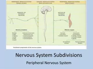
Dural venous sinuses • Venous channels found between layers of dura mater in the brain • They receive blood from internal and external veins of the brain, receive cerebrospinal fluid (CSF) from the subarachnoid space, and ultimately empty into the internal jugular vein • The walls of the dural venous sinuses are composed of dura mater lined with endothelium, a specialized layer of flattened cells found in blood vessels • They differ from other blood vessels in that they lack a full set of vessel layers (e.g., tunica media) • It also lacks valves as seen in veins
Inferior Sagittal Sinus • Occupies the free lower margin of the falxcerebri • Runs backward and joins great cerebral vein which is formed by the union of the 2 internal cerebral veins at the free margin of the tentoriumcerebelli to form the straight sinus • Receives cerebral veins from the medial surface of the cerebral hemisphere
Superior Sagittal Sinus • Occupies the upper fixed border of the falxcerebri • Begins in the front at the foramen cecum where it receives a vein from the nasal cavity • It runs backward, grooving vault of the skull and at the internal occipital protuberance it is continuous with the transverse sinus • It communicates through small openings with 2 or 3 venous lacunae on each side
Superior Sagittal Sinus • Numerous arachnoidvilli and granulations project into these lacunae which also receive the diploic; emissary and meningeal veins • It receives the superior cerebral veins • At the internal occipital protuberance it is dilated to form the confluence of the sinuses which is connected to the opposite transverse sinus and receives the occipital sinus
Straight Sinus • It occupies the line of junction of the falxcerebri with the tentoriumcerebelli • It is formed by the union of the inferior sagittal sinus with the great cerebral vein • It ends by turning to the left (sometimes to the right) to form the transverse sinus
Occipital Sinus • It is a small sinus occupying the attached margin of the falxcerebelli • It communicates with the vertebral veins near the foramen magnum • Superiorly it drains into the confluence of sinuses
Transverse Sinus • Paired and begin at the internal occipital protuberance • The right sinus usually continuous with the superior sagittal sinus • The left is continuous with the straight sinus • Each sinus occupies the attached margin of the tentoriumcerebelli , grooving the occipital bone and posteroinferior angle of the parietal bone • They receive the superior petrosal sinuses; inferior cerebral and cerebellar veins and diploic veins • They end by turning downward as the sigmoid sinuses
Superior and Inferior Prtrosal Sinuses • They are small and situated on the superior and inferior borders of the petrous part of the temporal bone on each side • Each superior sinus drains the cavernous sinus into the transverse sinus • Each inferior sinus drains the cavernous sinus into the internal jugular vein
Sigmoid Sinus • They are a direct continuation of the transverse sinuses • Each sinus turns downward and medially and grooves mastoid part of the temporal bone • It then turns downward through the posterior part of the jugular foramen to become continuous with the superior bulb of the internal jugular vein
Cavernous Sinus • Situatedin the middle cranial fossa on each side of the body of the sphenoid bone • Each sinus extends from the superior orbital fissure in front to the apex of the petrous part of the temporal bone behind • The 3rd ; 4th cranial nerves and the ophthalmic & maxillary divisions of the trigeminal nerve run forward in the lateral wall of this sinus • They lie between the endothelium and the dura mater • The internal carotid artery, its sympathetic nerve plexus and abducent nerve run forward through it • They are separated from the blood by an endothelial covering
Cavernous Sinus • The tributaries are: • 1- Superior ophthalmic vein which communicates it with the facial V • 2- Inferior ophthalmic vein. • 3- Cerebral veins • 4- Central vein of the retina • 5- Sphenopareital sinus. • The sinus drains posteriorly into the superior and inferior petrosal sinuses and inferiorly into the pterygoid venous plexus • The 2 sinuses communicate with one another by means of the anterior and posterior intercavernous sinuses which run in the diaphragmasellae in front and behind the stalk of the hypophysiscerebri
Cavernous Sinus • This diagram points out the structures found within the cavernous sinus and within its walls • In the walls: • 1 oculomotor • 2 trochlear • 4 V1 • 5 V2 • Within: 3 abducens • 6 autonomic plexus • 7 internal carotid artery • 8 pituitary gland 9 body of sphenoid bone
Veins of Brain • Delicate venous drainage from the cerebral hemispheres emerges from the brain to form small venous structures in the pia mater • These larger venous channels then form cerebral veins, which bridge the subarachnoid space and enter into endothelial-lined sinuses within the dura mater • Possess no valves • Have extremely thin walls • Pierce the arachnoid membrane and the inner or meningeal layer of the dura mater, and open into the cranial venous sinuses • Divided into two sets: • Cerebral • Cerebellar
Veins of Brain Divided into • External group(Superior, middle and inferior cerebral veins) • Internal group • Superior cerebral veins: Drain into the superior sagittal sinus • Middle cerebral vein: Drains in the cavernous sinus • Connected: • (a) with the superior sagittal sinus by the great anastomotic vein of Trolard, which opens into one of the superior cerebral veins • (b) with the transverse sinus by the posterior anastomotic vein of Labbé, which courses over the temporal lobe. • Inferior cerebral vein: Drain in the superior sagittal sinus, cavernous, sphenoparietal, and superior petrosal sinuses
Veins of Brain • Internal Cerebral Veins • Drain the deep parts of the hemisphere • Two in number • Formed near the interventricular foramen by union of Terminal vein and choroid veins • They run backward parallel with one another, between the layers of the telachorioidea of the third ventricle, and beneath the splenium of the corpus callosum, where they unite to form a short trunk, the great cerebral vein; just before their union each receives the corresponding basal vein • Great Cerebral Vein • Formed by the union of two internal cerebral veins • It is a short median trunk which curves backward and upward around the splenium of the corpus callosum and ends in the anterior extremity of the straight sinus
Arachnoid Mater • It forms a loose investment for the brain • Connected by delicate connective tissue with both the dura and pia mater • It surrounds the nerves, forming tubular sheaths for them as far as their points of exit from the skull. Unlike the pia mater, it does not dip into the sulci or fissures between the convolutions, but passes directly from one convolution to the other, bridging over the sulci • It is continued downward over the spinal cord • Because it is a serous membrane, it is a smooth, polished membrane to the naked eye
Arachnoid Mater • The delicate arachnoid layer is attached to the inside of dura and surrounds the brain and spinal cord • Cerebrospinal fluid (CSF) flows under the arachnoid in the subarachnoid space • The arachnoid mater makes arachnoidvilli, small protrusions through the dura mater into the venous sinuses of the brain, which allow CSF to exit the sub-arachnoid space and enter the blood stream
Pia Mater • Thin fibrous tissue impermeable to fluid • This allows the pia mater to enclose cerebrospinal fluid • By containing this fluid the pia mater works with the other meningeal layers to protect and cushion the brain • Allows blood vessels to pass through and nourish the brain • The perivascular space created between blood vessels and pia mater functions as a lymphatic system for the brain • When the pia mater becomes irritated and inflamed the result is meningitis
Pia Mater • The thin membrane is composed of fibrous tissue, which is covered by a sheet of flat cells impermeable to fluid on its outer surface • A network of blood vessels travels to the brain and spinal cord by interlacing through the pia membrane • These capillaries are responsible for nourishing the brain • This vascular membrane is held together by areolar tissue covered by mesothelial cells from the delicate strands of connective tissue called the arachnoidtrabeculae • In the perivascular spaces, the pia mater begins as mesothelial lining on the outer surface, but the cells then fade to be replaced by neuroglia elements
Pia Mater • Firmly adhered to the surface of the brain and loosely connected to the arachnoid layer • Because of this continuum, the layers are often referred to as the piaarachnoid or leptomeninges • A subarachnoid space exists between the arachnoid layer and the pia, into which the choroid plexus releases and maintains the cerebrospinal fluid (CSF) • The subarachnoid space contains trabeculae, or fibrous filaments that connect and bring stability to the two layers, allowing for the appropriate protection from and movement of, the proteins, electrolytes, ions, and glucose contained within the CSF
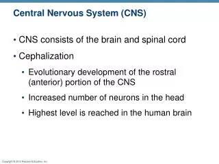
Pia Mater • In conjunction with the other meningeal membranes, pia mater functions to cover and protect the (CNS), to protect the blood vessels and enclose the venous sinuses near the CNS, to contain the (CSF) and to form partitions with the skull • The CSF, pia mater, and other layers of the meninges work together as a protection device for the brain, with the CSF often referred to as the fourth layer of the meninges
Pia Mater • Cerebrospinal fluid is circulated through the ventricles, cisterns, and subarachnoid space within the brain and spinal cord • About 150 ml of CSF is always in circulation • The CSF is primarily secreted by the choroid plexus, however about one-third of the CSF is secreted by pia mater and other ependymal surfaces of the ventricles and arachnoidal membranes • The ependymal surface refers to the thin epithelial membrane lining the brain and spinal cord canal • The CSF travels from the ventricles and cerebellum through three foramen in the brain, emptying in to the cerebrum, and ending its cycle in the venous blood
Arachnoid Granulations • Microscopic projections of the arachnoid into some of the venous sinuses • Prolongations of pia-arachnoid that protrude through the meningeal layer of the dura mater and have a thin limiting membrane • Collections of arachnoidvillus form arachnoid granulations that lie in venous lacunae at the margin of the superior sagittal sinus
Subarachnoid space • Space between arachnoid and pia mater • Occupied by spongy tissue consisting of trabeculae (delicate connective tissue filaments that extend from the arachnoid mater and blend into the pia mater) and intercommunicating channels in which the cerebrospinal fluid is contained
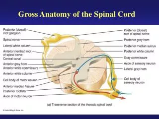
Dura Mater of Spinal Cord • The spinal cord, like the brain, is surrounded by the three meninges • The dura mater extends from foramen magnum to the sacrum and coccyx • The dura is attached to the foramen magnum and the periosteium covering the uppemost cervical vertebrae and their ligaments • Through the remainder of the vertebral canal, the dura is not attached to the vertebrae, being separated by the epidural (or peridural or extradural) space, which contains fat and the internal vertebral venous plexus • In caudal analgesia, an anesthetic solution injected into the sacral hiatus diffuses upward into the epidural space • This may be used in surgical procedures relating to pelvic and perineal regions • Extensions of dura (dural sheaths) surround the nerve roots and spinal ganglia, and continue into the connective tissue coverings (epineurium) of the spinal nerves
Arachnoid Mater of Spinal Cord • The arachnoid invests the spinal cord loosely • Continuous with the cerebral arachnoid above, it traverses the foramen magnum and descends to about the S2 vertebral level • The subarachnoid space, which contains cerebrospinal fluid (C.S.F.), is a wide interval between the arachnoid and pia • Because the spinal cord ends at about the level of the L2 vertebra, whereas the subarachnoid space continues to S2, access can be gained to the C.S.F. by inserting a needle between the vertebral lamina below the end of the cord, a procedure termed lumbar puncture • By this means, the pressure of C.S.F. can be measured, the fluid can be analyzed, a spinal anesthetic can be introduced, or fluid can be replaced by a contrast medium for radiography (myelography)
Arachnoid Mater of Spinal Cord • The arachnoid mater of the spinal cord is a thin, veil-like membrane between the dura mater and the pia mater • The arachnoid mater in the spinal cord is more delicate than the arachnoid of the brain, but it resembles it in sending tubular prolongations along the nerves • It is attached posteriorly to the dura mater by prolongations of connective tissue • Below, it is prolonged upon the caudaequina • The arachnoid mater forms a long sac, the cavity of which lies between the arachnoid mater and the pia mater, and is known as the subarachnoid space
Pia Mater of Spinal Cord • The pia mater invests the spinal cord closely, ensheathes the anterior spinal artery (as lineasplendens), and enters the anterior median fissure • Laterally, the pia forms a discontinuous longitudinal septum, the denticulate ligament, which sends about 21 tooth-like processes laterally to fuse with the arachnoid and dura on each side • The ligament is a surgical landmark in that it is attached to the spinal cord about midway between the attachments of dorsal and ventral roots