Download
1 / 42
430 likes | 678 Views
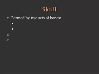
Pediatric Skull Xray. Heather Patterson August 2, 2007. Objectives. Brief review of anatomy Approach to pediatric skull xray Examples. Skull fractures. Common in non-accidental trauma 80% in first year Rare after 2y of age. Anatomy. Anatomy. Skull Xray.

E N D
Pediatric Skull Xray Heather Patterson August 2, 2007
Objectives • Brief review of anatomy • Approach to pediatric skull xray • Examples
Skull fractures • Common in non-accidental trauma • 80% in first year • Rare after 2y of age
Skull Xray • Full series 3-4 views • AP • Towne’s view (AP with neck flexed) • Lateral x 2
Approach • Follow cortex • Identify suture lines • Identify abnormal lines
What is the big deal? • Risk of “growing fracture” • Leptomeningeal cysts • Long term sequelae
Growing fracture/Leptomeningeal Cyst • Rare • <1% of skull fractures • Pathophys • Dural deal with herniation of pia and arachnoid through tear • CSF pulsations lead to erosion of bone • Diastasis of fracture over time
Growing fracture/Leptomeningeal Cyst • Imaging • Angular, linear lytic lesion • Scalloped margins • Management • f/u with neurosurgery • Early intervention as • needed
Case 1 • Linear fracture R posterior parietal and occipital bones • Extends through lambdoid suture
Case 2 • R parietal skull fracture
Case 3 • Linear fracture R occiput
Case 4 • Depressed skull fracture posterior right parietal bone
Case 5 • R parietal fracture • Communicates with lamboidal suture
Case 6 • R parietal fracture
Case 7 • L parietal fracture
Case 8 • Persistent skull defect • Encephalomalacic cystic defect • Consistent with leptomeningeal cyst