Download
1 / 32
320 likes | 327 Views
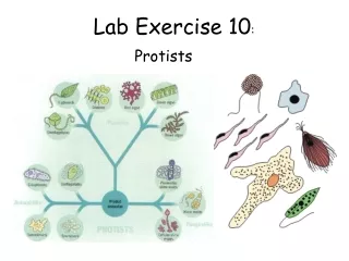
Learn about the anatomy of the spinal cord and spinal nerves, as well as different reflexes and the concept of learning. Activities include identifying structures, eliciting reflexes, and understanding reaction time and learning.

E N D
Lab 10 Anatomy of Spinal Cord and Spinal Nerves, Reflexes, and Reaction Time and Learning Joseph R. Schiller, Ph.D., James F. Thompson, Ph.D., and Gilbert Pitts, Ph.D.
Lab 10 Activities Identify spinal cord structures on microslides Identify spinal cord structures on models and charts, including the protective coverings Identify major nerve plexuses on nerve tree model (cervical, brachial, lumbar, + sacral) Elicit somatic, superficial, and autonomic reflexes in members of your lab group to learn about the various kinds of reflexes (stretch reflex, Babinski, pupillary + ciliospinal) Biopac Lesson 11 – Reaction Time and Learning: understand a basic difference in reflexes versus learning
Structure of the Spinal Cord • Gray Matter: deep “H” shaped region • White Matter: peripheral (myelinated) fiber tracts • Protective coverings: bone, meninges, related tissues • related structures: spinal nerve roots, spinal nerves, dorsal root ganglia
Functional Map of Gray Matter • Posterior (dorsal) (gray) horns: sensory axonal endings synapse with interneurons • Anterior (ventral) (gray) horns: somatic motor neuron cell bodies • Lateral (gray) horns: visceral = autonomic motor neuron cell bodies
Spinal Cord, c.s., silver stain, 40x • Silver stain produces sharp contrast between white and gray matter • Note the meninges and a dorsal root ganglion are also present
Spinal Cord, c.s., Masson, 40x • This slide also provides good contrast • Posterior/dorsal horn • Gray commissure • Central canal • Anterior/ventral horn
Spinal Cord, c.s., Thoracic, 40x • Gray commissure • Lateral horn
Lining of the Central Canal • Ependymal cells • in central canal
31 Pairs of Spinal Nerves • All are mixed (m/s) nerves • Thousands of fibers per spinal nerve • Each pair serves a particular region of the body, but overlaps some with the region supplied by the spinal nerve above and below it (redundancy)
Spinal Nerve Anatomy • Formed from junction of dorsal and ventral roots • Divide into: • dorsal ramus - supplies posterior body trunk • ventral ramus - supplies the rest of body trunk and the limbs
Branches of Ventral Ramus • Rami Comminicantes (autonomic system: supplies viscera) • White ramus communicans (to sympathetic chain) • Gray ramus communicans (from sympathetic chain) • Intercostal nerve (somatic motor and somatosensory supply to muscle and skin) • Cutaneous branches (supply skin)
Dermatomes • Areas of skin innervated by the cutaneous branches of each pair of spinal nerves • Each pair also provides some service to the region of the spinal nerve above and the spinal nerve below (redundancy)
Reflexes • A rapid, predictable, automatic response to a stimulus • Unlearned, unpremeditated, and involuntary • One is aware of somatic reflexes only after they occur • A homeostatic mechanism (feedback path) • Two fundamental types: • Somatic – effector is skeletal muscle • Autonomic (visceral) – effector is smooth muscle, cardiac muscle, or glands • Generally not consciously perceived
Components of a Reflex Arc • Receptor - dendrites or other sensory structures respond to changes in the internal or external environment • Sensory neuron - conducts from a receptor to axon terminals • Integrating center (region within the CNS) • Simple - monosynaptic (2 cells only: sensory and motor neurons) • Complex – polysynaptic (> 2 cells: interneurons involved) • Motor neuron - impulses from integrating center to effector • Effector - body part (muscle or gland) which responds to the motor nerve impulse
Stretch Reflexes • Receptor - muscle spindles • Mechanoreceptors which respond to stretching • Stimulus: stretch, causes increased nerve impulses to the spinal cord • Response: muscle contraction which reduces stretching of the muscle spindle and decreased nerve impulses to spinal cord
Stretch Reflexes • Remember, if a muscle is being stretched, its antagonist is contracting • This sensory proprioception information contributes to maintaining proper muscle tone
Patellar Reflex • Monosynaptic • Ipsilateral (same side) • Segmental (at one level of the spinal cord) • Reciprocal component – polysynaptic, inhibition of the antagonist
Golgi (Deep) Tendon Reflex • Receptor: Golgi tendon organ • Mechanoreceptor that responds to muscle tension (via the tendon) • Stimulus: increased tension (increased nerve impulses to spinal cord) • Response: muscle relaxes (decreased nerve impulses to spinal cord) • Inhibits the agonist • Reciprocal path: activates the antagonist • Polysynaptic, ipsilateral, and segmental
Flexor Reflex • A pull on the limb, extending it, will trigger the reflex • Also a painful stimulus – a burn, pin prick, toe stub, etc. • F-R causes an automatic withdrawal from the (dangerous) stimulus • Polysynaptic, ipsilateral, and segmental
Classifying Reflexes • Reflexes are classified according to: • Number of synapses in path: mono- versus polysynaptic • Location of receptor versus effector: ipsi- versus contralteral • Level of receptor versus effector: segmental (at same level) versus intersegmental (at different levels) • Patellar (stretch) reflex: monosynaptic, ipsilateral, segmental • Golgi Tendon Reflex: polysynaptic, ipsilateral, segmental
Crossed Extensor Reflex • Flexion of a body part is often balanced by extension of the same body part on the opposite side of the body • Polysynaptic • Contralateral • Segmental
Reflexes to Observe in Lab • Patellar or Knee Jerk Reflex • Ankle Jerk Reflex • Plantar (Babinski) Reflex – sole of the foot • Abdominal Reflex – if dressed appropriately • Pupillary Reflex • Ciliospinal Reflex
Biopac Lesson 11 • Reaction Time and Learning • Note: change font size to 9 before printing collected data • Learning requires the ability to connect one event (symbol, stimulus, pattern) with another • Reflexes do not • This lesson involves two different types of events: • Random: cannot be predicted and connected • Regularly repeated: can be learned and anticipated
segment 1 segment 2 segment 3 segment 4 Reformat Follow the onscreen instructions to collect five segments of data. Reformat the lesson-file name-day/date-time lines into a single line. Print your data journal with font size set to 8). You do not have to print the graph. Use the first four data segments to answer the questions from your lab guide on p. 10-21.
Lab 9 Written Homeworkto Turn in Next Week • Answer the questions and fill out the Table on p. 10-21 • Answer the questions on p. 10-23 • Attach your Biopac Data Journal • Put your name, your instructor’s name, day and time of your lab on all three pages