Download
1 / 17
231 likes | 601 Views
Lab no. 10. Plasmid DNA isolation. Purpose :. To learn how to extract plasmid DNA from E.coli . To observe the analysis of plasmid DNA by gel electrophoresis. Introduction :. Over the past decades it became evident that virtually in all bacterial species plasmids exist.

E N D
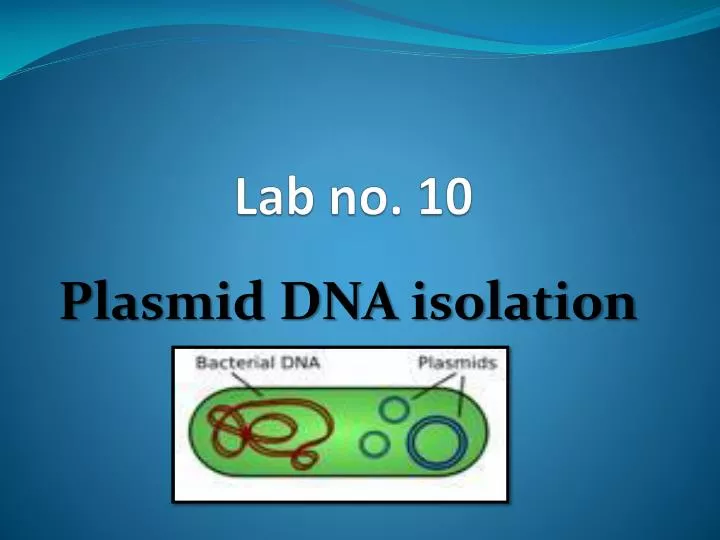
Lab no. 10 Plasmid DNA isolation
Purpose : • To learn how to extract plasmid DNA from E.coli. • To observe the analysisof plasmid DNA by gel electrophoresis.
Introduction : • Over the past decades it became evident that virtually in all bacterial species plasmids exist. • These accessory genetic elements are defined as autonomously replicating extra chromosomal DNA.
Plasmid • Genetic element not contained within a chromosome. • It occurs in many bacterial strains. • Plasmids are circular DNA molecules that replicate independently of the bacterial chromosome. • They are not essential for the bacterium but may give it a selective advantage. • Some plasmids determine the production of proteins that can kill other bacteria; others make bacteria resistant to antibiotics. • Plasmids are extremely valuable tools in the fields of molecular biology and genetics, specifically in the area of genetic engineering .
Plasmids typically account for only a small fraction of a bacterial genome corresponding roughly to a range between 1 and 200 kilobase pairs. However, extremely large megaplasmids with sizes far beyond 200kb have also been detected such as in Rhizobium and others. Plasmids of more than 50 kb might be characterized as large plasmids whereas plasmids used as tools in molecular genetics are often smaller than 10 kb.
Plasmid DNA can be separated from the rest of the bacteria DNA because it is much smaller circles than the circular bacterial chromosome. • Most procedures involve cell lysis in which the bacterial membrane is perforated and the contents released. • In the process, the long circular chromosome become sheared into linear fragments but the smaller plasmids remain intact. • Intact plasmid can be separated from linear DNA because they remain supercoiled.
The E. coli bacterial cells are resuspended in buffer containing glucose, Tris, EDTA, and RNase. Glucose : gives osmotic shock that leads to the rupture of cell wall and membrane. EDTA : stabilizes the cell membrane by binding divalent cations (Mg++ and Ca++) and EDTA is an important substance in all plasmid preparations because it inhibits nuclease activity. Rnase : destroys the cell’s RNA. The resuspended cells are then treated with a SDS and sodium hydroxide NaOH.
SDS dissolves the phospholipid and protein components of the cell membrane. • The cell membrane lyses, releasing the cell contents. • Sodium hydroxide denatures both plasmid and chromosomal DNA into single strands. • Chromosomal DNA separates completely into individual strands.
The single-stranded plasmid loops remain linked together like interlocked rings. • Acidic potassium acetate forms an SDS/lipid/protein precipitates. • Acetic acid neutralizes the NaOH. • At neutral pH, genomic DNA renatures and is trapped in the SDS/lipid/protein precipitate. • The plasmid DNA renatures into double-stranded molecules that remain in solution.
Procedure: • Grow up a 3 ml overnight of E. coli broth media. • Pour 1.5 ml into an eppendrof tube and Pellet cells by centrifuging at 12,000 rpm for 2 minutes. • Pour off supernatant and Add another 1.5ml of culture to the same tube and centrifuge 2 min and pour off the supernatent. • Resuspend the pellet by briefly vortex in 250µl of solution I(stored at 4ºC ).
Add 250 µl of solution II (stored at RT). Mix GENTLY by inverting and rotating the tube several times. DO NOT vortex. Put on ice 5 minutes. Add 350 µl of cold solution III (stored at 4 º C). Mix by inverting the tube 6-8 times. Put on ice for 5 minutes. Spin 12 minutes at 12000 rpm in a microcentrifuge at 4º C. (Pellet contains: SDS, Lipids, Proteins and Chromosomal DNA). Transfer 350 µl of supernatant (contains plasmid DNA). to a fresh tube.
Add 2 volumes ethanol at room tempreture. Mix and stand for 2min at RT. Pellet 5 min in microcentrifuge at RT. Dry, then resuspend in 50 µl TE. Analyze your preparations using agarose gel elctrophoresis by mix 8µl of the plasmid sample with 2µl of loading buffer or store at –20oC.