Download
1 / 67
820 likes | 1.39k Views
SKELETAL SYSTEM: VERTEBRAL COLUMN. Mrs. Ofelia Solano Saludar Department of Natural Sciences University of St. La Salle Bacolod City. PRIMARY COMPONENTS OF VERTEBRATE SKELETON :.

E N D
SKELETAL SYSTEM: VERTEBRAL COLUMN Mrs. Ofelia Solano Saludar Department of Natural Sciences University of St. La Salle Bacolod City
PRIMARY COMPONENTS OF VERTEBRATE SKELETON: Cartilage and bone, with additional support coming from fibrous (collagenous) connective tissue
These structural materials must be able to: • support the mass of the body and all of the muscles and organs that are part of the body • withstand tremendous forces that affect an organism • remain strong under the stresses of locomotion, such as when the feet strike the ground, sending the force of the impact through the body frame • be strong at the junction where two bones meet, where stress is applied and felt • protect against impact to soft tissues, such as the skull protecting the brain
In order to be strong, yet lightweight, bones are hollow in the middle Arc should minimize stress on the skeleton where the body mass is concentrated
CARTILAGE • A firm but elastic skeletal tissue • Matrix contains chondroitin sulfate (ground substance) and collagenor elastic protein fibers that bind with water • WHY ARE THERE NO CARTILAGINOUS TETRAPODS?
FORMS OF CARTILAGE HYALINE CARTILAGE- with a clear translucent matrix; found primarily on the ends of ribs and on the trachea ELASTIC CARTILAGE- contains elastin fibers that appears yellowish; found primarily on external ear and epiglottis
FIBROCARTILAGE contains collagen fibers; found in the intervertebral disks and pubic symphysis CALCIFIED CARTILAGE contains deposited calcium salts; found in the vertebrae of cartilaginous fish
BONE ORGANIC COMPONENT- primarily collagen, which gives bone its great tensile strength. INORGANIC COMPONENTS- largely calcium hydroxyapatitecrystals; comprise 60% of the dry weight and provide the compressive strength of bone.
BONE HISTOLOGY
Osteoblastsproduce new bone Osteoclastsremove and resorb existing bone by secreting acid to break down mineral component of bone and enzymes to break down the collagen component of bone Osteocytesmaintain equilibrium in fully formed bone Bone cells are identified based on their activity:
BONE FUNCTIONS • Provides support and movement via attachments for soft tissue and muscle • Protects vital organs • Is a major site for red marrow for production of blood cells • Plays a role in the metabolism of minerals such as calcium and phosphorus.
STRUCTURAL BONE TYPES: COMPACT BONEforms the outer shell of all bones and also the shafts in long bones. SPONGY BONEis found at the expanded heads of long bones and fills most irregular bones.
INTRAMEMBRANOUS OSSIFICATION • Bone formation begins with ablastema(any aggregation of pluripotent embryonic mesenchymal cells) • These cells then develop into either FIBROBLASTS (collagen) or OSTEOBLASTS (bone cells) . • Together, these form MEMBRANE BONE (bone deposited directed in a blastema). • Intramembranous ossification gives rise to: • Bones of the lower jaw, skull, and pectoral girdle • Dentin & other bone that develops in the skin • Vertebrae in some teleosts, urodeles, & apodans
ENDOCHONDRAL OSSIFICATION Process in which bone is deposited in pre-existing cartilage. Such bone is called REPLACEMENT BONE.
SKELETAL ELEMENTS 1. DERMAL SKELETON Ostracoderms had so much dermal bone they were called 'armored fishes‘ The skin of most living vertebrates has dermal bone elements usually present in the head region Tetrapods - retain dermal elements in the skull, jaws, & pectoral girdle
DERMAL BONE OF FISHES The evolutionary 'trend‘: large bony plates giving way to smaller, thinner bony scales
2. ENDOSKELETON a. VISCERAL SKELETON - associated with gills & skeletal elements (jaw cartilages) derived from them
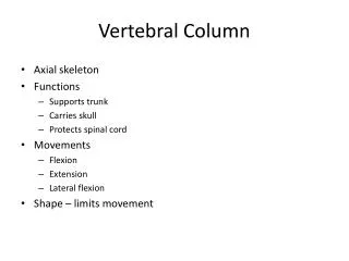
b. SOMATIC SKELETON Axial skeleton (vertebral column, ribs, sternum, & skull) + Appendicularskeleton
The endoskeleton develops from sclerotome, mesenchymethat accumulate around skeletogenous regions, the arrangement of which is largely dependent on the disposition of the myotomes. Myotomes are divided by myosepta, and divided into epaxial(dorsal), and hypaxial(ventral) halves by the horizontal septum. Around the notochord and neural tube, this septum continues middorsally as the dorsal septum, and midventrally as the ventral septum.
In the trunk region, this septum is divided into 2 ventrolateral septa by the intervention of the coelom. At the intersection of every myoseptum with the dorsal, ventral, and horizontal septa and with the perichordalmesenchyme, a vertebra arises. As the myosepta are segmented, the vertebrae are also segmented.
Sclerotomes of 2 adjacent segments become divided into a loose anterior half, and denser posterior half. The posterior halves of a pair of sclerotomes become the anterior part of a vertebra, and the anterior halves of the next pair become the posterior part of the same vertebra. As a result, the centrum of a vertebra intersects a myoseptum, and the vertebrae alternate with the myotomes.
The vertebral column originates as metamerically arranged endoskeletal elements aligned along the notochord and flanking the spinal cord. In agnathans, these vertebral elements or ARCUALIA, are only the basidorsals and the interdorsals. In the gnathostomes, there are additional ventral elements, the basiventrals and interventrals.
Mesenchyme from sclerotomes accumulate around the notochord to form the perichordalmesenchyme. • The notochord and its sheath may calcify into centra.
Basidorsals and basiventrals come from the original posterior halves of the sclerotomes, form the neural and hemal arches, and contribute to the centrum. Interdorsals & interventrals come from the original anterior halves of the sclerotomes. Interdorsals give rise to the intercalary arch, or contribute to the neural arch or centrum. Interventrals are of less importance. Primitive vertebrae are composed of separate pieces; a stage before components have fused.
In primitive amniotes, 2 pieces appeared: the pleurocentrumcorresponding to the interdorsalarcualia, and the hypocentrum, the basiventrals. The pleurocentrum forms the main mass of the centrum; the hypocentrum remains as hemal arches (chevron bone) in the tail, and also contributes to the atlas and sometimes the axis.
The ensemble produced by the calcified notochord & sheath, plus fusions of the arcualia contributes to the formation of the centrum& archesof the vertebra. • Vertebrae compose the vertebral column.
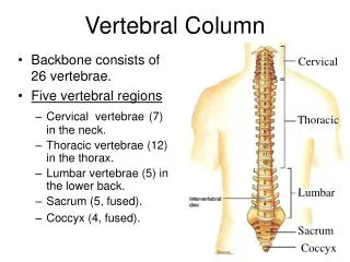
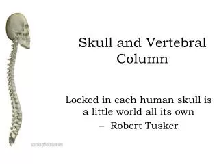
VERTEBRAL COLUMN Vertebrae - consist of a centrum (body), 1 or 2 arches, plus various processes
Acelous - flat-ended; mammals Amphicelous - concave at both ends; most fish, a few salamanders (Necturus), and caecilians Procelous - concave in front and convex in back ; anurans and present-day reptiles Opisthocoelous - convex in front & concave in back; most salamanders Heterocelous - saddle-shaped centrum at both ends; birds TYPES OF CENTRA
Vertebral Processes: projections from arches & centra; some give rigidity to the column, articulate with ribs, or serve as sites of muscle attachment • Transverse processes - most common type of process; extend laterally from the base of a neural arch or centrum & separate the epaxial & hypaxial muscles • Diapophyses & parapophyses- articulate with ribs • Prezygapophyses(cranial zygapophyses)& postzygapophyses (caudal zygapophyses) - articulate with one another & limit flexion & torsion of the vertebral column
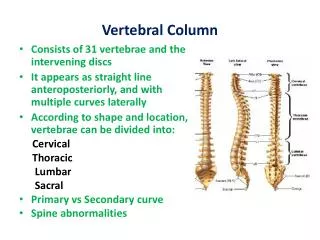
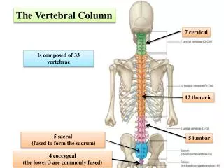
Dorsal Region • Dorsals- name given to vertebrae between cervicals & sacrals when all articulate with similar ribs (e.g., fish, amphibians, & snakes) • Crocodilians, lizards, birds, & mammals - ribs are confined to anterior region of trunk • thoracic – 12-15 vertebrae with ribs • lumbar - vertebrae without ribs
CERVICAL region Atlas - 1st cervical vertebra; ring-like (most of centrum gone); provides 'cradle' in which skull can 'rock' ; hypocentrum; pleurocentrum is odontoid process of axis Axis- 2nd cervical vertebra; 2 pleurocentra Transverse foramen (#6) found in cervical vertebrae provides canal for vertebral artery & vein
SACRAL vertebrae • Have short transverse processes that brace the pelvic girdle & hindlimbs against the vertebral column • Sacrum - single bony complex consisting of fused sacral vertebrae; found when there is more than 1 sacral vertebra
CAUDAL region • Primitive tetrapods - 50 or more caudal vertebrae • Present-day tetrapods - number of caudal vertebrae is reduced, arches & processes get progressively shorter (the last few caudals typically consist of just cylindrical centra
PHYLOGENY OF VERTEBRAL COLUMNS • Vertebral column of the earliest tetrapods did not consist of 1 bone/body segment. • Crossopterygian vertebrae consisted of an hypocentrum plus 2 pleurocentra. This type of vertebra is called a rachitomous vertebra. • The 'trend' in vertebrate evolution has been for pleurocentrato increase in size, and for the hypocentrumto decrease in size.
Agnathans - only skeletal elements associated with the notochord are paired, lateral neural cartilages • Cartilaginous fishes do not have typical fish vertebral columns. Vertebrae include neural arches (dorsal plates of cartilaginous arcualia) and dorsal intercalary plates are located between successive arches
Teleosts • Regional differentiation: trunk and caudal regions • Well-ossified amphicelous vertebrae • Centra form from perichordal and sheath elements
Chondrosteans (sturgeons & paddlefish) & modern lungfishes • Incomplete centra • Notochord is not constricted • Cartilage deposited in notochord sheath provides structural support
Basidorsal and basiventrals only • Notochord persists within each centrum (but constricted) • Neural arch associated with each centrum and hemal arches in tail (caudal) vertebrae
Diplospondyly = 2 centra and 2 sets of arches per body segment; occurs in some fish (including sharks)
Amphibians • Regional differentiation: • Cervical (1 vertebra, limited head movement) • Trunk(ribs present) • Sacral(1 vertebra) • Caudal(no zygapophyses) • Basidorsals appear; basiventrals reduced • Centra is perichordalmesenchyme represented by hypocentrum; pleurocentrum form intervertebral disks • Craniovertebral joint with 1-2 occipital condyles • Urostyle– fused post-sacral vertebrae in anurans basiventrals)
TETRAPODS • Terrestrial life: spine must support body, resist bending but provide mobility • Regional differentiations: cervical, thoracic, lumbar, sacral, caudal
Centrum is the descendant of the pleurocentrum • Vertebra compounded of arch and perichordal components