Download
1 / 20
300 likes | 664 Views
SALIVARY GLANDS. Dr. Jameela El- Medany. OBJECTIVES. By the end of this lecture the student should be able to : Describe the anatomy of the parotid gland: position, shape, structures within it , innervation and parotid duct.

E N D
SALIVARY GLANDS Dr. Jameela El-Medany
OBJECTIVES By the end of this lecture the student should be able to: Describe the anatomy of the parotid gland: position, shape, structures within it ,innervation and parotid duct. Describe the anatomy of the submandibular and sublingual salivary glands: location, shape, parts, ducts and innervation of the glands.
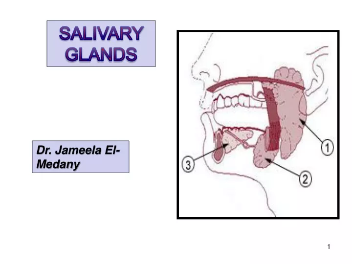
Salivary glands are exocrine glands, that produce saliva. There are 3 large named pairs of salivary glands and multiple minute unnamed glands in the submucosa of the oral cavity(lips, palate & under surface of the tongue). Parotidproduces a serous, watery secretion. Submandibular produces a mixed serous & mucous secretion. Sublingualsecretes saliva that is predominantly mucousin character. ndibular
Largestsalivary gland. Formed entirely of serous acini. Position: Wedged between mandibular ramus & masseteranteriorly, Mastoid process & sternomastoid muscle posteriorly PAROTID GLAND
Triangular: Apex behind angle of the mandible Base directed upward just below the zygomatic arch, external auditory meatus &TMJ. Accessory part: A small part that is separated from the main gland. SHAPE
Capsule: Tight, derived from deep cervical fascia of the neck. The gland is divided into superficial & deep parts, by the facialnerve fibers.
Parotid Duct It opens into the vestibule of the mouth on a small papilla, opposite the upper second molar (maxillary) tooth.
What are the Structures within the Parotid gland? From superficial to deep 1- Facial nerve: It is the most superficial structure, it divides the gland into superficial & deep parts. 2- Retromandibular vein:intermediate in position Formed by the union of maxillary & superficial temporal veins. Before it leaves the gland it is divided into anterior & posterior branches. 3- External carotid artery:Most deep, It isdivided into maxillary and superficial temporal arteries.
Gives: TWO Branches before it enters the gland FIVE Branches within the parotid: 1- Temporal 2- Zygomatic 3- Buccal 4- Mandibular 5- Cervical. FACIAL NERVE
Nerve Supply: Parasympatheticfrom inferior salivary nucleus – tympanic nerve- through the glossopharyngeal nerve to tympanic plexus- lesser petrosal to otic ganglion- The postganglionic fibers running in auriculotemporal nerve. Sympathetic: from plexus around external carotid artery.
Blood supply Arterial:ECA & its branches. Venous drainage: retromandibular vein. Lymphatic: parotid & deep cervical lymph nodes.
SUBMANDIBULAR SALIVARY GLAND Located deep to the body of the mandible
PARTS • Formed of 2 parts: • Large superficial part • Small deep part
SUBMANDIBULAR DUCT • The duct emerges from the deep part of the gland. • It passes forward along the side of the tongue, under the mucous membrane of the floor of the mouth. • It is crossed laterally by the lingual nerve • It opens on the summit of a small sublingual papilla, which lies at the side of the frenulum of the tongue.
SUBMANDIBULAR DUCT • Clinically, it is important to remember that the submandibular duct can be palpated through the floor of the mouth alongside the tongue. • Saliva can usually be seen emerging from the orifice of the duct.
CALCULUS FORMATION • The submandibular duct is a common site of calculus formation. • The presence of a tense swelling below the body of the mandible, which is greatest before or during a meal and is reduced in size or absent between meals, is diagnostic of the condition. • Examination of the floor of the mouth will reveal absence of ejection of saliva from the orifice of the duct of the affected gland. • Frequently, the stone can be palpated in the duct, which lies below the mucous membrane of the floor of the mouth.
SUBLINGUAL GLAND LOCATION • The smallest of the three salivary glands. • It lies below the mucous membrane of the floor of mouth, close to the midline.
Sublingual ducts • The sublingual ducts are 8 to 20 in number. • Most open into the summit of the sublingual fold, but a few may open into the submandibular duct.
Blood Supply Arterial supply: Facial artery. Venous drainage: Facial vein. Lymph drainage: Submandibular lymph nodes.
NERVE SUPPLY • Parasympathetic secretomotor supply is from superior salivary nucleus of the facial (7th) nerve. The fibers pass to the submandibular ganglion via the chorda tympani nerve and the lingual nerve. • Postganglionic parasympathetic fibers reach the submandibular & sublingual glands either directly or along the duct.