Download
1 / 102
1.04k likes | 1.13k Views
Diseases of Salivary Glands. Diseases of salivary gland : Developmental anomalies Sialadenitis Obstruction and traumatic lesions SjÖgren syndrome Sialadenosis HIV-associated salivary gland disease Salivary gland tumour Age changes in salivary gland. 1- Developmental anomalies.

E N D
Diseases of salivary gland : • Developmental anomalies • Sialadenitis • Obstruction and traumatic lesions • SjÖgren syndrome • Sialadenosis • HIV-associated salivary gland disease • Salivary gland tumour • Age changes in salivary gland
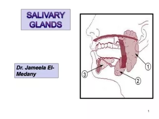
1- Developmental anomalies • Aplasia of one or more major glands and atresia of one or more major salivary gland ducts. • Congenital aplasia of the parotid gland may be associated with other facial abnormalities e.g mandibulofacial dysostosis, aplasia of the lacrimal gland and hemifacial microsomia. • Heterotopic salivary tissue in a variety of sites in head and neck, most frequent at the angle or within the body of the mandible ( present as stafne’s idiopathic bone cavity). • Accessory parotid tissue within the cheek or masseter muscle.
2- Inflammatory disorders Sialadenitis Inflammatory disorders of the major salivary gland are usually result of bacterial or viral infection but occasionally due to other causes such as trauma, irradiation and allergic reactions.
Sialadenitis Sarcoidosis Bacterial Postirradiation Viral Acute Sialadenitis of minor gland Mumps Chronic Cytomegalic inclusion disease
A- Acute Bacterial Sialadenitis • Uncommon disorder principally involve the parotid gland. • Acute parotitis is an ascending infection ( Bacterial reach the gland from the mouth by ascending the ductal system).
Etiology : Streptococcus pyogenes and staphylococcus aureus , less common Haemophilus species or members of the black-pigmented bacteroides group. • Reduced salivary flow is the major predisposing factor. • Acute parotitis may occur in patients with sjogren syndrome or following the use of drugs with xerostomic side-effect. • Acute infection may arise in immunocompromisedpatients or as result of acute exacerbation in a previously chronic sailadenitis.
Clinical feature: • The onset is rapid. • Swelling accompanied by pain , fever , malaise and redness of overlying skin. • Pus may be expressed from effected duct.
B- Chronic bacterial sailadenitis • Non-specific inflammatory disease associated with duct obstruction ( due to salivary calculi) and low grade ascending infection. • The submandibular gland more common than parotid.
Clinical feature: • - Usually, unilateral and symptoms of recurrent tender swelling of the effected gland to associated obstruction. • The duct orifice appear inflamed. • Purulent or salty-tasting discharge ( in acute exacerbations ).
Histological examination: • Dilatation of the ductal system. • Hyperplasia of duct epithelium. • Periductal fibrosis and acinar atrophy with replacement fibrosis. • Chronic inflammatory cell infiltration. - In sialography may see the duct obstruction, destruction of glandular tissue and duct dilatation ( sialectasia ).
- Progressive chronic inflammation in submandibular gland may result in almost complete replacement of the parenchyma by fibrous tissue producing a firm mass that may be mistaken clinically for a neoplasm.
Recurrent parotitis: • Is a rare disorder may effect children or adults. • Rarely the adults form following from childhood type, due to persistence of factors (calculi or duct strictures) leading to recurrent attacks of low-grade ascending infection.
The aetiology of childhood type is unclear • Low secretion rate predisposing to ascending infection. • Immaturity of the immune response in infants. • Congenital abnormalities of the ductal system.
Clinical feature: • Unilateral or bilateral. • Recurrent painful swelling of the gland. • Pus may be expressed from the duct orifice. • In most cases the condition resolvesspontaneously by the time but repeated infection may result in irreversible damage to the main duct lead to duct obstruction then lead to ascending infection and damage then recurrent parotitis in adult life.
B- Viral Sialadenitis: • Mumps(epidemic parotitis ) • It is the commonest cause of parotid enlargement and the commonest of all the salivary gland diseases. • Is an acute, contagious infection, which often occurs in minor epidemics.
Etiology: Caused by a paramyxovirus. • The virus is transmitted by direct contact with infected saliva and by droplet spread. • Incubation period of 2-3 weeks.
Clinical feature: • - None-specific prodromal symptoms of fever and malaise followed by sudden onset of painful swelling in one or more salivary glands (parotid glands are always involved bilaterally in about 70% of cases. • Occasionally the submandibular and sublingual gland may be affected (rarely without parotid involvement) • The gland enlargement gradually subsides in about 7 days.
The virus is present in the saliva 2-3 days before the onset of disease and for about 6 days afterwards. • Occasionally, in adult other internal organs are involved such as testes, ovaries, central nervous system and pancreas (orchitis is common complication is 20% of cases in adult males). • Recurrent of infection is rare (long lasting immunity).
Diagnosis: • Is usually made on clinical grounds. • In atypical cases can be confirmed by the detection of , IgM, antibodies and in serum antibody titer to mumps virus antigens.
B- Cytomegalic inclusion disease • (Salivary Gland inclusion disease) • Infection with cytomegalovirus (member of the herpesvirus group). • Most primary infection are asymptomatic but the virus can cause severe disseminated disease in neonates and immunocompromised host (such as transplant patients and HIV-infected patients). • Usually, incidental histological finding.
Presence of large, doubly contoured “owl-eye” inclusion bodies within the nucleus or cytoplasm of duct cells of the parotid gland. • Similar inclusion frequently occur in the kidneys, liver, lungs, brain and other organs.
C- Postirradiation sialadenitis: -is common complication of radiotherapy and there is a direct correlation between the dose of irradiation and the severity of the damage. -if the damage is severe, it would be irreversible leading to fibrous replacement of the damage acini and squamous metaplasia of ducts. -in less severe damage, some degree of function may return after several months. -serous acini are more sensitive to radiation damage than mucous acini.
D- Sarcoidosis: -systemic chronic granulomatous disorder of unknown aetiology. -most common effects young adults. -present most frequently with bilateral hilar lymphadenopathy , pulmonary infiltration and skin or eye lesion. -oral mucosa involvement is rare(incident onset, signs and symptoms disappear in time but some time leave residual swelling. -they may effect the parotid and minor salivary gland.
- parotid involvement present as persistent, often painless, enlargement and may be associated with involvement of the lacrimal glands in heerfordt syndrome or uveoparotid fever ( sarcoidosis with combination of uvitis, parotitis and facial paralysis).
E- Sialadenitis of minor salivary gland : -incidental and insignificant finding. -it is seen most frequently in association with mucous extravasation cysts and stomatitis nicotina of the palate. -very rarely, it is present with multiple mucosal swellings associated with cystic dilatation of ducts and chronic suppuration, this condition referred to (stomatitisglandularis). It occur most commonly on the lips.
Allergic sialadenitis • Treatment: - Self-limited disease - Supportive therapy - Avoid allergen - Hydration
3-Obstructive and traumatic lesions Causes: 1-due to a blockage within the lumen- sialolith 2-result from disease in or around the duct wall such as fibrosis or neoplasia. 3-obstruction to the duct orifice is due to chronic trauma such as sharp cusps or over-extended denture resulting in fibrosis and stenosis.
A-salivary calculi (sialoliths): -it is cause obstruction within the duct lumen. -common in middle-aged adults. -submandibular gland 70-90% of cases parotid gland sublingual and minor gland is uncommon. - deposition of calcium salts around an initial organic nidusaltered salivary mucins, desquamated epithelial cells , microorganisms. -calculi are usually unilateral, may form in ducts within the gland or in the main excretory duct, multiple stone in the same gland are not uncommon.
-the typical sign and symptom are pain and suddenenlargement of the gland, especially at meal time when salivary secretion is stimulated. -the reduction of salivary flow( predispose to ascending infection and chronic sialadenitis). -the calculi my detected by palpation and on radiograph, and may be round or ovoid, rough or smooth, and vary in size. -they are usually yellowish in color and composed from calcium phosphates and smaller amount of carbonates.
Sialolithiasis Treatment • Conservative: good hydration, antibiotics and anti-inflammatory, milking of gland spontaneous stone passage. • Excision: - - Lithotripsy - Sialendoscopy • Transoral removal- manipulation fails then a surgical cut is made into duct • Gland excision - the stone is within the gland - the gland is severely damaged by chronic infection.
B- Necrotizing sialometaplasia: -is a relatively uncommon disorder which clinically and on histological examination may be mistaken for malignant disease.
Aetiology: Is unknown, but ischaemia leading to infarction of salivary lobules is the most accepted theory. In some patient there may be history of trauma from a variety of causes, including local anesthetic injection and previous surgery.
Clinically • It occur most frequently on the hard palate in middle-aged patient ,and is about twice as common in men as women. • -it present as a deep crater-like ulcer which may mimic a malignant ulcer and take up to 10-12 weeks to heal.
Histopathological examination: -lobular necrosis of salivary gland, squamous metaplasia of ducts and acini , mucous extravasation, and inflammatory cell infiltration. -the overlying palatal mucosa show pseudoepitheliomatous hyperplasia and the histopathological feature may be mistaken for either squamous cell carcinoma or mucoepidermoid carcinoma.
3- SJÖGREN SYNDROME: Is chronic autoimmune disease characterized by lymphocytic infiltration and acinar destruction of lacrimal and salivary glands leading to dry eyes (keratoconjunctivitis sicca ) and dry mouth (xerostomia). Classification: 1-primary sjÖgren syndrome: combination of dry mouth and dry eye. 2-secoundary sjÖgren syndrome: occurs in association with another autoimmune connective tissue disease, most frequently rheumatoid arthritis or systemic lupus erythematosis (this type occur in about half of the cases).
Aetiology: The specific cause of this syndrome is unknown, Multifactorialprocess, with immunological alteration indicating a disease of great complexity. - sjÖgren syndrome occurs with increased frequency in patient with HLA class 11 genes and several virus especially Epstein-barr virus.
>Clinical features: -predominantly affects middle-aged female (female : male ratio is about 9:1) - drynessand soreness of mouth and eyes. -xerostomiadifficulty in swallowing and speaking ,increased fluid intake -Xerostomia oral candidiasis, bacterial sialadenitis, dental caries and periodontal disease . -oral mucosa appear dry, smooth and glazed.
-tongue: the dorsum of the tongue appears red and atrophic with varying degree of fissuring and lobulation. -salivary gland enlargement (in 30% of patient) , this enlargement is bilateral, parotid gland most common and is seldom painful. -keratoconjunctivitis sicca manifests as dryness of the eyes with conjunctivitis and cause gritty and burning sensation.