Download

1 / 54
540 likes | 669 Views
How I manage pulmonary nodular lesions and nodular infiltrates in patients with hematologic malignancies or undergoing hematopoietic cell transplantation.

E N D
How I manage pulmonary nodular lesions and nodular infiltrates in patients with hematologic malignancies or undergoing hematopoietic cell transplantation
Various case series suggest that 13% to 60% of patients develop a pulmonary infiltrate at some point in their treatment course, with the incidence varying considerably by different diseases and treatments.
High-resolution CT (HRCT) scans are the preferred method for evaluation of a lung infiltrate over radiography because they are more sensitive in detecting infiltrates earlier and they are more capable of better characterization of the infiltrates.
In patients treated for acute leukemia or undergoing HCT, HRCTscanshave a high degree of sensitivity (85%), high negative predictive value (85%) in detecting pneumonia,
Clinical management is often changed because of the HRCT findings.
pulmonary infiltrates can be categorized by their radiographic pattern broadly into diffuseand nodular infiltrates.
Nodular lesions may be further characterized as solitary micronodulesor macronodules with sharp or unsharpmargins with or without halos, multiple nodules, masses, nodular infiltrates, and focal airspace disease.
Both infectious and noninfectious etiologies can be the cause of nodules and nodular infiltrates.
Hodgkin or non-Hodgkin lymphoma and much less so for patients with leukemia • plasmacytomasas extramedullary manifestations of multiple myeloma • case reports of acute myelogenousleukemia (AML)
More commonly, pulmonary infiltrates in patients newly presenting with AML are diffuse and may result fromleukostasisin those with high leukocyte counts or even frank leukemic infiltration of tissues, pulmonary hemorrhage, and less commonly infection.
Patients newly presenting with AML occasionallypresent with pulmonary infections.
The types of infections that cause nodular infiltrates before start of chemotherapy have not been well described but, in our experience, are mostly bacterial. Both Gram-positive organisms (especially staphylococci and streptococci) and Gram-negative organisms (especially Pseudomonasaeruginosa, Escherichia coli, and Klebsiella spp.) are common.
Fungal pneumonia is infrequent in newly diagnosed AML at initial presentation and, where seen, occurs predominantly in those with antecedent cytopenias or iron overload.
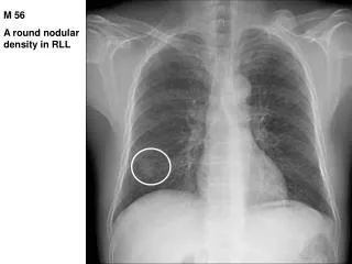
Nodules in patients not highly immunosuppressed or myelosuppressedmay also be caused by the same types of processes that cause pulmonary nodules in nonimmunosuppressed patients. In noncompromisedpatients, approximately half of nodules are caused by malignancy, chiefly primary lung cancer (usually solitary nodules) or less commonly metastases (usually multiple nodules).The other half are mostly the result of infectious granulomata from mycobacteria or fungi and much less commonly from other infrequent benign etiologies, such as hamartomas, sarcoidosis, or arteriovenousmalformations.
Infections account for most nodular infiltrates in patients receiving active chemotherapy or immunotherapy or have severe compromise in immunity. Bacterial and fungal infections most commonly account for nodularinfiltrates, whereas viruses, Pneumocystisjiroveciand Legionellamost commonly account for diffuse infiltrates.
Bacterial pneumonia during neutropenia can be caused by both Gram-positive and Gram-negative organisms. Early after HCT, staphylococcusand Gram-negative pathogens are problematic,
Invasive fungal infections, especially by mold pathogens, are particularly common, especially in patients with deep prolonged neutropenia during AML therapy. The onset is frequently later during neutropenia, typically occurring beyond 2 weeks of neutropenia.
Approximately 45% of nodular infiltrates in AML treatment are the result of aspergillosis. In our experience, at least half of nodular infiltrates in AML therapy and HCT are the result of fungi, with 80% of the pulmonary fungal infections caused by aspergillosis, the remainder resulting from other molds, such as the agents of mucormycosis, and less frequently, fusarium, and Scedosporium species.
It is also important to recognize that nodular lesions may be the result of more than one organism. Mixed mold infections (most commonly aspergillosis and mucormycosis) can occur. In addition, aspergillosiscan be accompanied by coinfection by bacteriaor viruses. This growing recognition of mixed infections emphasizes the need for establishing a specific diagnosis.
Noninfectious causes must also be kept in mind, although they are less common. Noninfectious conditions include primary lung cancer, metastases from other epithelial cancers, lymphomas, EBV-associated posttransplantation lymphomas after HCT, and pulmonarythromboembolism. Rare conditions, such as Wegener granulomatosis, sarcoidosis, and pulmonary arteriovenousmalformation, should also be considered.
How do I make the diagnosis? • Can Imaging distinguish various etiologies?
The HRCT scan is the best imaging technique to evaluate pulmonary infiltrates. A variety of studies in nonimmunocompromisedpatients have noted size, location, calcification pattern, change in size over time, edge characteristics, internal characteristics, number of nodules, attenuation, and contrast enhancement as features that provide important information.
Lesions less than1 cm are infrequently the result of neoplasm. Larger nodules are more likely to be malignant. Masses (lesions 3 cm) are highly likely to be malignant. Malignant lesions are more likely to be in the upper lobes, whereas nonmalignant etiologies are more evenly distributed. Calcification is very suggestive of a benign granulomatous etiology if it has an organized diffuse, central, or laminar pattern.
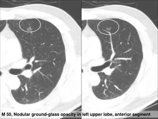
Lesions stable for more than 2 years are rarely malignant. • Spiculated edges are suspicious for malignancy. • Satellite nodules surrounding a central larger nodule are suggestive of granulomatous disease. • An air-bronchogram within a nodule is suggestive of malignancy. • Cavitation may be seen in both malignant and nonmalignant entities. • Ground-glass opacificationis suggestive of malignancy, such as adenocarcinoma in situ or minimally invasive adenocarcinoma of the lung.
Using such considerations, in noncompromised patients, lesions can categorized as demonstrating low, indeterminant, or high probability of malignancy. Those lesions judged to be high risk should undergo resection. Those at low risk (small 8 mm, benign calcification pattern, stable over time, no smoking history) can be followed over time.Thosejudged to be indeterminantrequire further evaluation.
PET scans have been shown to be quite useful in the evaluation of the solitary pulmonary nodule (SPN) in the nonimmunocompromisedpatient. PET scans may be of particular value in lesions of indeterminantsignificance because malignancies typically have high uptake
PET scans have been found to be useful in lymphoma patients to distinguish active disease from inactive scar. However, PET has not been found to be useful for small lesions less than 1 cm because of false-negative results. • Those at high risk require resection or biopsy. • Infectious etiologies may also have high signal uptake by PET.
A number of studies have evaluated imaging findings as they relate to specific etiologies in patients treated for HM or undergoing HCT. • For bacterial pneumonias, airspace consolidation is most common, but small centrilobular nodules and ground-glass opacities are also common; large nodules also are seen but are less common (20%). The halo sign appears to be the most useful radiologic sign for distinguishing aspergillosis from bacteria.
In patients with invasive aspergillosis (IA), one large study, in mostly HM and HCT therapy, found that 94% had one or more macronodules(at least 1 cm in diameter). In 79% of cases, the nodules were multiple, and in 60%, there were multiple bilateral nodules. Halo signs (dense nodules surrounded by ground-glass perimeter) were found in 61%. Other findings noted with IA were consolidation(30%), infarct-shaped nodules (27%), cavities (20%), and air-crescentsigns (10%).
The nodular lesions of IA are often peripherallylocated. The presence of dense, well-circumscribed nodule, air-crescent sign, or cavity has been adopted as specific radiologic criteria that along with appropriate clinical setting and with supporting microbiology criteria establish the diagnosis of IA in consensus guidelines. In earlier studies, serial HRCT scans were performed in patients with IA: halo signs were most likely to be found early in infection, air-crescent signs much later,typically found at time of neutrophil recovery.
Although IA is the most likely cause of halo lesions in this population, it is important to note that the halo is now known to not be specific for IA: other pathogens, such as Paeruginosa, the agents of mucormycosis, and other less common molds can also give rise to halo infiltrates.
Mucormycosis can present with pulmonary nodules or nodular infiltrates similar to IA. There may be some distinguishing findings on CT scan. Comparing IA and mucormycosis, the presence of more than 10 nodules, pleural effusion, or sinusinvolvement, and a history of prior voriconazole use (an antifungal active against Aspergillusspecies but not mucormycosis) were conditions more likely to be found with mucormycosis than with IA.
Important to note is that, although pulmonary nodules are highly likely in IA, other less-specific radiologic manifestations can also be caused by IA, including consolidation, ground-glass infiltrates, and occasionally pleural effusion. The reversed halo (circular focus of ground-glass density within a ring of dense consolidation) was initially described as highly suggestive of mucormycosisand subsequently also IA, but other infectious and noninfectious etiologies may also present with this radiologic picture.
In studies of patients with AML who were neutropenicand patients who underwent HCT with lung opacities more than 5 mm, both IA and bacterial pneumonia were manifest frequently by both nodules and air-space consolidation in similar proportions.Thehalo sign was rarely seen in bacterial pneumonia, but cavities an air-crescent signs were present in both. A recent study suggests a role for CT pulmonary angiography in the diagnosis of pulmonary mold infections, exploiting the fact that such infections are angioinvasive.
Can other noninvasive techniques be helpful? • Gram stain and culture of sputum should be performed if sputum is being expectorated; unfortunately, the patient who is neutropenicis frequently unable to expectorate sputum. Bacterial and fungal blood cultures should be performed; when positive, they are helpful; however, most have negative blood cultures. Mycobacterial blood cultures should also be considered in the evaluation of nodular lesions in non-neutropenic patients.
There are 2 commercial serum assays to assist in the diagnosis of invasive fungal infections. The serum galactomannan (GM) and Beta -glucantests are useful in detection of invasive fungal infections. It is important to note that the test is recommended to be performed twice weekly prospectively in patients at risk, and the sensitivity and specificity of a single test result drawn at the time of a lung infiltrate are less.Neither of these fungal serologic tests can detect the agents of mucormycosis.
Even given the limitations of the 2 commercial serum fungal assays, we recommend their use. When positive, we launch an investigation in search of further confirmation of an invasive fungal infection. Even when negative, if either the clinical scenario and/or imaging suggest fungal pneumonia, we recommend proceeding to invasive techniques in search of an invasive fungal infection or to establish a specific alternative diagnosis.
Invasive techniques to establish the diagnosis • However, FB with bronchoalveloar lavage (BAL) is a common approach for evaluation of nodular and diffuse infiltrates in HM and HCT patients.The presence of symptoms, location more centrally, presence of bronchus sign on CT, and visualization during bronchoscopy are associated with higher yields.
Use of noncultural microbial testing can also increase the yield of BAL. The use of BAL GM testing has been associated with substantially increased yield in patients with IA. In a meta-analysis of BAL GM, summary estimates of the BAL-GM assay for proven or probable IA were as follows: sensitivity, 90%; specificity, 94%; The estimates of the BAL GM assay for provenIA were as follows: sensitivity, 94%; and specificity, 79%.82
In our view, the greater safety of FB along with comparable yield has shifted the preference from TTNA and surgical lung biopsy to FB in most situations in the highly immunosuppressed HM and HCT setting, particularly where infection is most likely.
Moreover, the delay in establishing a diagnosis not covered by the presumptive therapy is likely to result in poorer treatment outcome because delays are associated with lower responses. Further, delayed investigation is associated with lower diagnostic yields. A study of early (within 4 days of presentation) versus late bronchoscopy in HCT patients found a 2.5-fold higher yield compared with later bronchoscopy and greater mortality in patients subjected to late FB. The yield was highest (75%) when bronchoscopy was performed within 24 hours of presentation. For these reasons, we urge performance of an invasive procedure at presentation rather than waiting to determine response to initial therapy, pursuing an invasive diagnostic only in those who are not responding
How should I assess the response totherapy? • For patients with small lesions at low risk for active infection or malignancy in which the initial decision was to observe, additional scanning at 2 to 3 months is advisable.
For those who were found to have an active infection, response to therapy dictates the type and frequency of additional subsequent testing. If clinically responding, repeat imaging should be done periodically until the infection is resolved. It is important to note that infiltrates may take several weeks to 1 to 2 months to fully resolve; thus, radiology by itself should not dictate the need for further diagnostic interventions.
It is well recognized that the infiltrates in patients with IA worsen over the first week of therapy, even though with continued therapy, the patients respond. The patient with IA who is not clinically improving is the most challenging situation.