Download
1 / 21
260 likes | 338 Views
Dr. Eman El Eter. Physiology of Consciousness. Objectives. Levels of consciousness/ definition Functional divisions of RF. Overview of functions of RF. Anatomical components of RAS. Connections of RAS. Neurotransmitters of RAS. Functions of RAS. What is Consciousness ?.

E N D
Dr. Eman El Eter Physiology of Consciousness
Objectives • Levels of consciousness/ definition • Functional divisions of RF. • Overview of functions of RF. • Anatomical components of RAS. • Connections of RAS. • Neurotransmitters of RAS. • Functions of RAS.
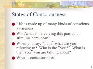
What is Consciousness ? • Is the brain state in which a person is being aware of the self and surroundings . • It is a product of electrical activity of the brain • (a person with a flat EEG can not be conscious ! )
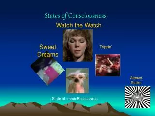
What are the levels of consciousness? • (1) Normal Consciousness (state of normal arousal , being fully awake and aware of the self and surroundings ) • (2) Clouded consciousness : person conscious but mentally confused ( e.g., in cases of drug or alcohol intoxication , high fever associated with malaria or septicemia , dementia , etc ) . • (3) Sleep : person unconscious ( in relation to the external world & surroundings ) , but is arousable ( can be aroused ) . • (4) Coma : person unconscious and not arousable
What are brain Structures involved in the conscious state? Consciousness depends upon interactions between : (1) Reticular Formation ( RF) . (2) Thalamus (3) Cortical Association areas .
Reticular formation • This regulates many vital functions including the sleep/awake cycle. It is a polysynaptic network located in the pons, midbrain and upper medulla and is poorly differentiated. It consists of 3 parts: • Lateral Reticular Formation • Has small neurones • Receives information from ascending tracts for touch and pain. • Receives vestibular information from median vestibular nerve. • Receives auditory information from superior olivary nucleus. • Visual information from superior colliculus. • Olfactory information via medial forebrain bundle
Reticular Formation, cont.,, • Paramedian Reticular Formation • Has large cells. • Receives signals from lateral reticular formation. • Projects onto cerebral hemispheres. • Nucleus coeruleus contains noradrenergic neurones and projects onto the cerebral cortex. • Ventral tegmental nucleus contains dopaminergic neurones that project directly onto the cortex. • Cholinergic neurones project onto the thalamus
Reticular formation, cont.,, • Raphe nuclei (Median RF) • In the midline of the reticular formation • Contain serotonergic projections to the brain and spinal cord.
What are the Functions of reticular formation? • 1. Somatic motor control (Reticulospinal tracts) • 2. Cardiovascular control - The reticular formation includes the cardiac and vasomotor centers of the medulla oblongata. • 3. Pain modulation - The reticular formation is one means by which pain signals from the lower body reach the cerebral cortex. It is also the origin of the descending analgesic pathways. The nerve fibers in these pathways act in the spinal cord to block the transmission of some pain signals to the brain.
Functions of RF, continued,…. • 4. Sleep and consciousness - The reticular formation has projections to the thalamus and cerebral cortex . It plays a central role in states of consciousness like alertness and sleep. Injury to the reticular formation can result in irreversible coma. • 5. Habituation - This is a process in which the brain learns to ignore repetitive, meaningless stimuli while remaining sensitive to others. A good example of this is when a person can sleep through loud traffic in a large city, but is awakened promptly due to the sound of an alarm .
Thalamus: The thalamus is contained in the mid-part of the diencephalon and is split up into a number of different nuclei. Cholinergic projections from the thalamus are responsible for: • Activation of the cerebral cortex. • Regulation of flow of information through other thalamic nuclei to the cortex via projections into reticular nuclei. • Tuberomammillary nucleus in the hypothalamus projects to the cortex and is involved in maintaining the awake state.
Anatomical components of RAS • The RAS is composed of several neuronal circuits connecting the brainstem to the cortex . These pathways originate in the upper brainstem reticular core and project through synaptic relays in the rostralintralaminar and thalamic nuclei to the cerebral cortex.As a result, individuals with bilateral lesions of thalamic intralaminar nuclei are lethargic or somnolent. • Several areas traditionally included in the RAS are: • Midbrain Reticular Formation. • Mesencephalic Nucleus (mesencephalon) • Thalamic Intralaminar nucleus • Dorsal Hypothalamus. • Tegmentum.
RAS • Lesion in the mid-pons makes the animal spends the rest of its life unconscious . • This means that areas in the upper pons and midbrain are essential for wakefulness . That area called BulboreticularFacilitory ( Excitatory ) Area of the reticular formation .
Functions of RAS:1- Regulating sleep-wake transitions • The main function of the RAS is to modify and potentiate thalamic and cortical functions resulting in (EEG) desynchronization. • Low voltage fast burst brain waves (EEG desynchronization) are associated with wakefulness and REM sleep , • During non-REM sleep, neurons in the RAS will have a much lower firing rate large voltage slow waves . • The physiological change from a state of deep sleep to wakefulness is reversible and mediated by the RAS. • Stimulation of the RAS produces EEG desynchronization by suppressing slow cortical waves. • In order that the brain may sleep, there must be a reduction in ascending afferent activity reaching the cortex by suppression of the RAS.
2-Attention • The reticular activating system also helps mediate transitions from relaxed wakefulness to periods of high attention.
3-RAS and learning • The RAS is the center of balance for the other systems involved in learning, self-control or inhibition, and motivation. • When functioning normally, it provides the neural connections that are needed for the processing and learning of information, and the ability to pay attention to the correct task.
What happens if RAS is not working properly? If RAS is depressed: * An under-aroused cortex, with difficulty in learning, poor memory, little self-control, and so on. *lack of consciousness or even coma. • If the RAS is too excited, and aroused the cortex or other systems of the brain too much? hyper-vigilance, touching everything, talking too much, restless, and hyperactive.
Indices of Level of Consciousness • Appearance & Behavior : • posture ( sitting , standing ? ) , open eyes ? . Facial expression ? , responds to stimuli ( including the examiner’s questions about name , orientation in time & place ? & other general Qs like who is the president ? ) • Vital signs : • Pulse , BP, respiration , pupils , reflexes , particularly brainstem reflexes , etc ) • EEG Each of these states ( wakefulness , sleep , coma and death ) has specific EEG patterns . • Evoked potentials ( in cases of Brain Death ).
Brain Death Confirmatory Testing with EEG Normal EEG ( at normal magnification ) Brain Death ( Flat EEG ,at very high magnification )
Brain Death Confirmatory Testing with Somatosensory Evoked Potentials Stimulation of a sense organ can evoke a cortical response that can be recorded by scalp electrode over the primary receiving cortical area for that particular sense .