Download
1 / 36
360 likes | 389 Views
Formation and differentiation of mesoderm, derivates of the germinal layers. Dr Gallatz Katalin. What happened until now?. FROM DR. MARK KOZSUREK. Cleavage of the zygote Consist of repeating mitotic divisions of the zygote, Resulting in a rapid incease of number of cells

E N D
Formation and differentiation of mesoderm, derivates of the germinal layers Dr Gallatz Katalin
What happened until now? FROM DR. MARK KOZSUREK
Cleavage of the zygote Consist of repeating mitotic divisions of the zygote, Resulting in a rapid incease of number of cells In this stage the dividing cells are called Blastomeres become smaller with each division. After 9-cell-stage: compaction mediated by adhesion glycoproteins. Morulafrom 12-16 cell stage inner and outer cell mass embryoblaststrophoblasts
3. Formation of the blastocyst Zona pellucida degenerates, fluid flows in, cavity appears between the cells blastocystic cavity
Summary of first week of the human development Stage 1. fertilization formation of the zygote Stage 2. 2-3. days early stage of cleavage morula Stage 3. 4-5. days free (unattached ) blastocyst Stage 4. 5-6. days attachment of the blastocyst IMPLANTATION
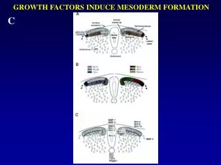
A TRILAMINARIS Stage – 3. week 1. The hypoblasts induce the epiblasts, 2. The epiblasts proliferate, 3. From the caudal side of the embrionic disc develops the primitive streak, at the end of it appears the primitive node and pit 4. Dividing cells migrate under the epiblast layer along the primitive streak , 5. Mesoderm is formed TRILAMINAR EMBRIO: Ectoderm , mesoderm , endoderm
Formation of mesoderm ( 3-8. week)
Formation of the notochord 1. Prenotochordalcellsinvaginatingintheprimitive pit moveforwardtotheprechordalplate 2. Notochordfuseswiththeunderlyingvisceralhypoblasts, and itsinferiorwall degenerates and disappear. 3. Neuroentericcanal is formedbetweentheamnion and yolksacfor a shorttime. 4. Definitív notochorddevelopsfromthecellsof notochordalprocessbetweenthe prechordalplate and cloacalmembrane Neuroenteric canal
MESODERM Paraxial mesoderma - somite Intermediate mesoderma gononephrotom Parietal mesoderm somatopleura splanchnopleura
DIFFERENTIATION OF THE MESODERM 17th day paraxial mesoderm lateral mesoderm . intermediate mesoderm (gononephrotom). Lateral mesoderm divides into 2 layers somatopleura and splanchnopleura. The cavity between them: intraembryonal coeloma. It is continouos with extraembryonal coeloma.
FORMATION OF THE SOMITES • At the beginning of the 3rd week somites (or somitomeres) appear both side of • the notochord and developing neural tube • Somites appear first cranially (around the 20th day), than develop caudally • (3 pair/day). • - 4 OCC, 7 CERV, 12 THOR, 5 LUMB, 5 SACR, 8-10 COCC • - In the head region neuromers (mesenchyma of the head) develop.
SOMITOGENESIS AND DIFFERENTIATION 4th week dermatomyotom SCLEROTOM Ventromedial cells of the somites migrate and surround the neural tube and notochordsclerotom vertebra The remaining part is called dermatomyotom myotom(muscles develop from it) dermatom, the dermis and subcutis develops from it
Somites are segmented in the whole length of the embryo, intermediate mesoderm is segmented rostrally only, while the lateral plate mesoderm is not segmented at all.
DERIVATES OF THE INTERMEDIATE MESODERM • - The intermediate mesoderma or gononephrotom gives the kidneys and • gonads • In the cervical and upper thoracic region it is segmentated , lower it • forms a continuos nephrogen cord
Derivates of the lateral mesoderm Fromthesomatopleuradevelopsthe body wall (PARIETAL LAYER OF THE SEROUS MEMBRANES). Fromthesplanchnopleuramesothelialorserousmembranesdevelop (pleura, pericardium, peritoneum).
neural tube somite intermediate mesoderm kidney, ureter, male genital tract notochord splanchnic mesoderm visceral layer of serous membranes gut lateral plate mesoderm somatic mesoderm parietal layer of serous membranes intraembryonic coelom→ body cavities (pericardium, pleura, peritoneum) FROM DR. MARK KOZSUREK
ectoderm epiblasts primitive streak intraembryonic mesoderm yolk sac endoderm hypoblasts ? endoderm In fact all the tissues and cells of the human are derived from the epiblasts! extraaembryonic mesoderm FROM DR. MARK KOZSUREK
EMBRYOGENESIS 1.Separation of theembrional-extraembrionaltissues 2. Gastrulation: formation of thegermlayers – EKTO-, ENDO-, MESODERM 3. Neurulation: formation of theneuraltube and crest (CNS AND PNS) 4. Differentiation of themesoderm
ECTODERM • Neuroectoderm neural plate neural crest placodes 2. Surface ectoderm FROM DR. MARK KOZSUREK
NEURAL PLATE – NEURAL TUBE Neurons and glial cells of the central nervous system CNS (except the microglia) Retina, dilator and constrictor of pupil muscles of the eye NEURAL CREST Peripheral nervous system PNS medulla of the adrenal gland melanocytes (which contain melanin pigment) aorticopulmonary septum of the heart odontoblasts
PLACODS otic placode: epithelial and neural elements of the vestibulocochlear system (organs of hearing and equilibrium) olphactory (nasal) placode: primary neuroepithelial cells of the nasal cavity optic (lens) placode: lens of the eye adenohypophyseal placode: hormone secreting cells of the adenohypophysis trigeminal placode:gives rise to the cells of the trigeminal ganglion epibranchial placode: taste buds of the tongue, soft palate and laryngeal inlet, neurons and supporting cells of taste-sensing ganglia of the facial glossopharyngeal and vagus nerves. SURFACE ECTODERM Epidermis, hair, nails, sebaceous and sweat glands of the skin Epithelium of the corne and conjunctiva, epithelium of the lacrimal apparatus Epithelium and glands of the oral and nasal cavity, enamel of the teeth, salivary glands Mammary glands Epithelium and glands of the anus, epithelium of the spongy part of the male urethra
MESODERM EPITHELIAL TYPE (densly packed cells) Intermediate mesoderm → epithelium of the kidney, ureter and the male genital tract (epididymis, ductus deferens, seminal vesicle) Somato-splanchnopleural junction →cortical cells of the suprarenal gland, gonadal epithelia (not including the germ cells!), epithelium of the uterine tube, uterus and the upper vagina Somato- and splanchnopleura→ mesothelium (simple squamous epithelium) MESENCHYMAL TYPE Circulatory system, including the endothelium of blood and lymph vessels, cardiac muscle, formed elements of blood and lymphatic cells Connective and supportive tissues – all of them!!! Smooth muscles (not including those in the eyeball!) Microglia
ENDODERM Epithelium and glands of theGI tract(includingliver, gallbladder and pancreas) Epithelium of theurinarybladder, theprostate, theprostatic part of themaleurethtraand thewholelength of thefemaleurethra Epithelium and glands of theairways(trachea, bronchi and alveoli of thelungs) Epithelium of thetympaniccavity and auditorytube Thyroid, parathyroidglands Thymus, tonsills