Download
1 / 26
290 likes | 474 Views
Immunoglobulins. Dr Nayyer uz Zaman Lecturer Biochemistry KGMC. Antibodies. Antibodies are globulin proteins ( Immunoglobulins ) that react specifically with the antigen that stimulated their production. They make up about 20% of the protein in blood plasma

E N D
Immunoglobulins DrNayyeruz Zaman Lecturer Biochemistry KGMC
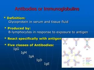
Antibodies • Antibodies are globulin proteins (Immunoglobulins) that react specifically with the antigen that stimulated their production. • They make up about 20% of the protein in blood plasma • Blood contains three types of globulins: alpha, beta and gamma • Antibodies are gamma globulins
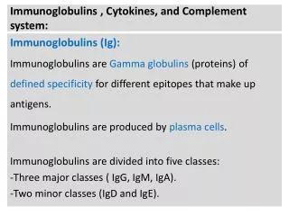
IMMUNOGLOBULINS • There are five classes of immunoglobulins (Antibodies): • IgG • IgA • IgM • IgE • IgD
Immunoglobulin Structure • Immunoglobulins are glycoproteins made up of LIGHT (L) and HEAVY (H) polypeptide chains • The terms light and heavy refer to the molecular weight • Light chains have a molecular weight of about 25,000 while heavy chains have a molecular weight of 50,000 to 70,000
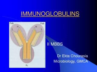
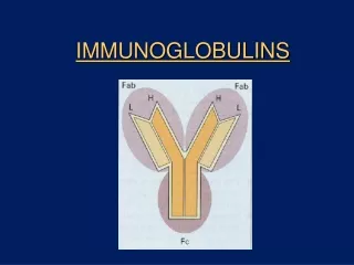
IMMUNOGLOBULINS • The simplest immunoglobulin molecule has a “Y” shape and is made up of four polypeptide chains: Two “H” chains and two “L” chains. • The four chains are linked by disulfide bonds • An individual antibody molecule always consists of two identical H chains and two identical L chains
Two Forms of Immunoglobulin Membrane-bound receptor Soluble antibody
IMMUNOGLOBULINS • L and H chains are sub-divided into “variable” and “constant” regions • The variable regions are responsible for antigen binding • The constant regions are responsible for various biological functions such as complement activation and binding to cell surface receptors
Immunoglobulin Structure Variable(V) & Constant (C) Regions VL & CL VH & CH Disulfide bond Carbohydrate CL VL CH2 CH3 CH1 Hinge Region VH
Humoral Immunity • Also known as antibody-mediated immunity • It is directed primarily against • Toxin induced diseases • Infections by encapsulated organisms (egpneumococcus, Haemophilus etc) • Certain viral infections
IMMUNOGLOBULINS • THE PRIMARY RESPONSE: • When an antigen is first encountered, the antibodies are detectable in the blood after a long lag period i.e. 7-10 days. • A small clone of B cells or plasma cells specific for the antigen is formed • The first antibodies to form are IgM, followed by IgG or IgA
IMMUNOGLOBULIN • THE SECONDARY RESPONSE: • When there is a second encounter with the same antigen, months or years after the primary response, there is a RAPID antibody response with HIGHER levels of immunoglobulins (antibodies) and a shorter lag period (only 3-5 days) • There is a much larger amount of IgG made than IgM, although IgM is first to be made in the secondary response. • This is due primarily to the “memory” B-cells that were formed during the primary response. • This is the basis for the concept of vaccination as well
Immunoglobulin Classes IgG • Each IgG molecule consists of two L chains and two H chains linked by disulfide bonds • There are 4 subclasses IgG1 – IgG4 (with IgG1 making up most i.e. approx 65% of total IgG) • IgG is the predominant antibody in the secondary immune response and constitutes and important defense against bacteria and viruses
IgG • IgG is the only antibody that can cross the placenta, therefore it is the most abundant antibody in newborns • IgG is one of two antibodies that can activate the complement system (IgM is the other one) • IgG is the immunoglobulin that opsonizes i.e. it can help enhance phagocytosis
IgA • IgA is the main immuoglobulin in secretions such as colostrum, saliva, tears and respiratory, intestinal and genital tract secretions • It prevents attachment of micro-organisms e.g. bacteria and viruses to mucous membranes.
IgM • IgM is the main immunoglobulin produced in the primary immune response. • It is present as a monomer on the surface of virtually all B-Cells where it functions as an antigen binding receptor. • It is a pentamer • It is the most efficient immunoglobin in agglutination, complement fixation, and other antibody reactions and is important in defense against bacteria and viruses.
IgM • It can be produced by the fetus in certain infections • Does not cross the placenta
IgD • This immunoglobulin has no known antibody function but may function as an antigen receptor • It is present on the surface of many B-lymphocytes • It is present in small amounts in serum
IgE • IgE is medically important for two reasons • It mediates immediate hypersensitivity (anaphylaxis) • It participates in host defenses against certain parasites, e.g. helminths (worms)
IgE • IgE (Fc portion) of IgE binds to the surface of mast cells and basophils. • Bound IgE serves as a receptor for antigens (allergen) and this antigen-antibody complex triggers allergic responses of the immediate (anaphylactic) type through the release of mediators e.g. histamine • Although it is present in trace amounts in normal individuals, its levels are raised in persons with allergies
IgE • IgE is the main host defense against certain important helminth (worm) infections, such as strongyloides, trichinella, ascaris and the hookworms. • The serum IgE level is usually increased in these infections. • Because worms are too large to be ingested by phagocytosis, they are killed by eosinophils that release worm-destroying enzymes
IgE • IgE specific for worm protiens binds to receptors on eosinophils, triggering the antibody-dependent cellular cytotoxicity (ADCC) response.
C-reactive protein (CRP) CRP is a major component of acute phase proteins . It is produced in the liver and is present in the circulation in minute concentration (< 1 mg/dl). C-creative protein (C strands for carbohydrate to which it binds on the capsule of pneumococi) is involved in the promotion of immune system through the activation of complement cascade. Estimation of CRP in serum is important for the evaluation of acute phase response. In a normal surgery, serum CRP increases and returns to normal level within 7-10 days. If the recovery is complicated by any infection, it will be reflected by the continuous elevation of CRP which requires further treatment.
Multiple Myeloma • Multiple myeloma, a plasma cell cancer constitutes about 1% of all cancers affecting the population. Females are more susceptible than mates for this disorder and it usually occurs in the age group 45-60 years. • Abnormal Ig production: Multiple myeloma is due to the malignancy of a single clone of plasma cells in the bone marrow . This results in the overproduction of abnormal immunoglobulins mostly (75%) IgG and in some cases (25%) IgA or IgM IgD type multiple myeloma. In patients of multiple myeloma, the synthesis of normal immunoglobulins is diminished causing depressed immunity. Hence recurrent infections are common in these patients. • Electrophoretic pattern: The plasma of multiple myeloma patients show a characteristic pattern of electrophoresis . There is a sharp and distinct band (M band , for myeloma globulin ) between ß-and γ-globulins. Further, this M band almost replaces the γ-globulin band due to the diminished synthesis of normal γ-globulins.
Bence jones proteins: Henry Bence Jones first described them in 1847. There are the light chains (k or γ) of immunoglobulins that are synthesized in exces. Bence Jones proteins have a molecular weight of 20,000 or 40,000 (for dimer). In about20./. Patients of multiple myloma. • Bence Jones proteins are excreted in urine which often damges the renal tubules. • 2. The classical heat test involves the precipitation of Bence Jones proteins when slightly acidified urine is heated to 40-50 0 C . This precipitate redissolves on further heating of urine to boiling point. It reappears again on cooling urine to about 700 C. • 3. Bradshaw`s test involves layering of urine on concentrated HCL that forms a white ring of precipitate if Bence Jones proteins are present.